. 2010 May 1;21(9):1609-19.
doi: 10.1091/mbc.e09-11-0923. Epub 2010 Mar 10.

Yeast Mpk1 cell wall integrity mitogen-activated protein kinase regulates nucleocytoplasmic shuttling of the Swi6 transcriptional regulator

Affiliations

Yeast Mpk1 cell wall integrity mitogen-activated protein kinase regulates nucleocytoplasmic shuttling of the Swi6 transcriptional regulator

Ki-Young Kim et al. Mol Biol Cell. .

Abstract

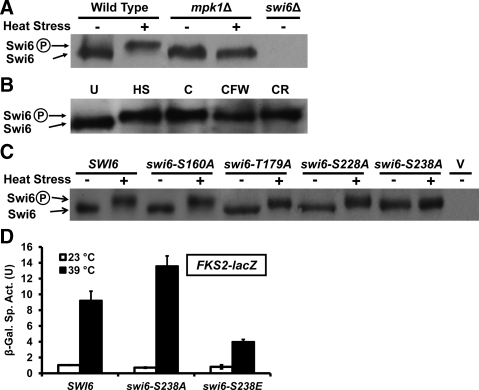
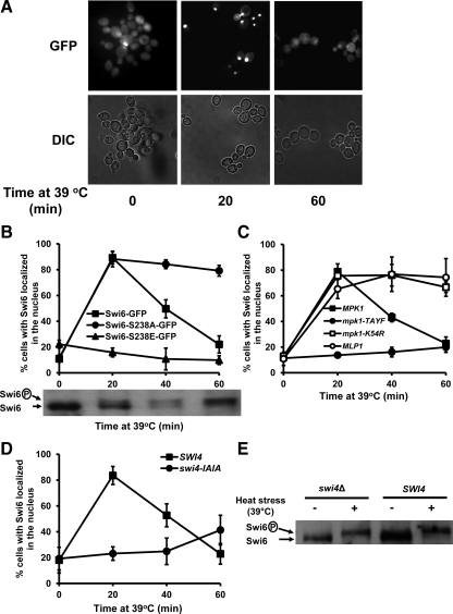
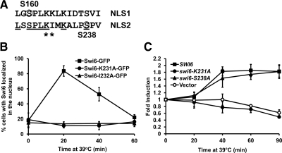
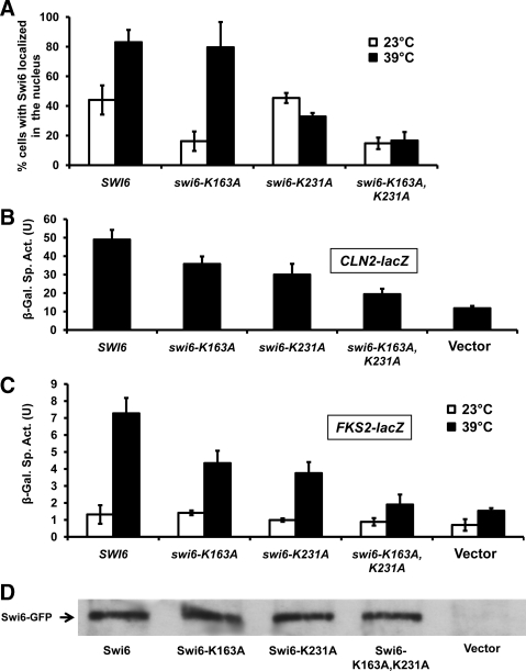
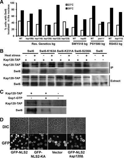
The yeast SBF transcription factor is a heterodimer comprised of Swi4 and Swi6 that has a well defined role in cell cycle-specific transcription. SBF serves a second function in the transcriptional response to cell wall stress in which activated Mpk1 mitogen-activated protein kinase of the cell wall integrity signaling pathway forms a complex with Swi4, the DNA binding subunit of SBF, conferring upon Swi4 the ability to bind DNA and activate transcription of FKS2. Although Mpk1-Swi4 complex formation and transcriptional activation of FKS2 does not require Mpk1 catalytic activity, Swi6 is phosphorylated by Mpk1 and must be present in the Mpk1-Swi4 complex for transcriptional activation of FKS2. Here, we find that Mpk1 regulates Swi6 nucleocytoplasmic shuttling in a biphasic manner. First, formation of the Mpk1-Swi4 complex recruits Swi6 to the nucleus for transcriptional activation. Second, Mpk1 negatively regulates Swi6 by phosphorylation on Ser238, which inhibits nuclear entry. Ser238 neighbors a nuclear localization signal (NLS) whose function is blocked by phosphorylation at Ser238 in a manner similar to the regulation by Cdc28 of another Swi6 NLS, revealing a mechanism for the integration of multiple signals to a single endpoint. Finally, the Kap120 beta-importin binds the Mpk1-regulated Swi6 NLS but not the Cdc28-regulated NLS.


Figures

Figure 1.
Figure 1.
Cell wall stress-induced phosphorylation of Swi6. (A) Mild heat shock induces an Mpk1-dependent band shift in Swi6. Wild-type yeast (1788), an mpk1Δ mutant (DL456), and a swi6Δ (DL3148) mutant were grown to mid-log phase in YPD + 2% sorbitol at 23°C and subjected to heat shock (39°C) for 2 h before extract preparation and immunoblot detection of Swi6. (B) Other cell wall stresses induce phosphorylation of Swi6. Wild-type yeast strain (1788) was grown to mid-log phase in YPD at 23°C and subjected to the indicated cell wall stress for 2 h before extract preparation and immunoblot detection of Swi6. U, unstressed; HS, heat shock (as in A); C, caffeine (8 mM for 2 h); CFW (40 μg/ml for 2 h); and CR, Congo red (50 μg/ml for 2 h). (C) Identification of Ser238 of Swi6 as a cell wall stress-induced phosphorylation site. A swi6Δ strain was transformed with centromeric plasmids expressing wild-type Swi6 (p2542), the indicated point mutants, or the parent vector (V; pRS313). Transformants were grown to mid-log phase in YPD at 23°C and subjected to heat shock as described above before detection of Swi6. (D) Effects of Swi6 phosphorylation site mutations on FKS2 transcription. An FKS2-lacZ reporter plasmid (p2052) was cotransformed with a centromeric plasmid expressing wild-type Swi6 (p2542), Swi6-S238A (p2547), or Swi6-S238E (p2548) into a swi6Δ strain (DL3233). Transformants were grown to saturation at 23°C in selective medium. Cultures were diluted into 3 ml of YPD so that subsequent incubation at 23 or 39°C for 15 h resulted in mid-log phase cultures (A600 of 1.0–1.5). β-Galactosidase activity was measured in crude extracts. Each value represents the mean and SD from three independent transformants. Sp. Act., specific activity; U, unit.
Figure 2.
Figure 2.
Nucleocytoplasmic shuttling of Swi6. (A) Representative fluorescent (GFP) and differential interference contrast (DIC) images of a swi6Δ mutant (DL3148) transformed with a centromeric plasmid expressing wild-type Swi6-GFP (p2391). Transformants were cultivated at 23°C in the presence of nocodozole to synchronize them in G2 phase before exposure to cell wall stress by heat shock for the indicated times. (B) Top, a swi6Δ mutant (DL3148) was transformed with plasmids expressing wild-type Swi6-GFP (p2391), Swi6-S238A-GFP (p2557), or Swi6-S238E-GFP (p2558). Cells were treated as described in A, except that populations of cells were scored visually for Swi6-GFP localization. Each value represents the mean and SD from three experiments in which at least 100 cells were scored. Bottom, a wild type strain (DL100) was treated as described above and subjected to immunoblot detection of Swi6. (C) An mpk1Δ mlp1Δ (DL3327) strain was cotransformed with centromeric plasmids expressing wild-type Mpk1 (p2188), Mpk1-T190A, Y192F (Mpk1-TAYF; p2190), Mpk1-K54R (p2193), or 2-μ Mlp1 (p2022), and wild-type Swi6-GFP (p2647). For this experiment, cells were treated as described in B, except that 2% sorbitol was added to the medium for osmotic support. (D) A swi4Δ mutant (DL3145) was cotransformed with centromeric plasmids expressing wild-type Swi6-GFP (p2647) and either wild-type Swi4 (p2713) or Swi4-I913A, I915A (Swi4-IAIA; p2714). Cells were treated as described in B. (E) A swi4Δ mutant (DL3145) was transformed with a centromeric plasmid expressing wild-type Swi4 (p2713) or vector (pRS315). Transformants were subjected to heat shock for 2 h, and cell extracts were processed for immunoblot detection of endogenous Swi6.
Figure 3.
Figure 3.
Swi6 possesses two NLS sequences. (A) Amino acid sequence alignment of Swi6 NLS1 and NLS2. Residues phosphorylated by Cdc28 (Ser160) and Mpk1 (Ser238) are circled. Asterisks indicate the residues (K231 or I232) mutated to Ala. (B) Mutations in NLS2 block CWI signaling-induced nuclear import of Swi6. A swi6Δ mutant (DL3148) was transformed with a centromeric plasmid expressing wild-type Swi6-GFP (p2391), or mutants in the putative NLS2 sequence, Swi6-K231A-GFP (p2730), or Swi6-I232A-GFP (p2731) and treated as in Figure 2 for assessment of Swi6 localization. (C) A mutation in NLS2 blocks FKS2 transcription in response to cell wall stress in cells arrested in G2. A swi6Δ mutant (DL3233) was transformed with a centromeric plasmid expressing wild-type Swi6 (p2542), mutants in the putative NLS2 sequence, Swi6-S238A (p2547), Swi6-K231A (p2832), or vector (pRS313). Transformants were treated as described in B, but FKS2 mRNA levels were measured by RT-PCR at the indicated time points and normalized against 18S RNA. Each value represents the mean and SD from three experiments.
Figure 4.
Figure 4.
Two NLS sequences in Swi6 display partial overlap of function. (A) Nuclear localization of Swi6 mutant forms in cycling cells. A swi6Δ mutant (DL3148) was transformed with a centromeric plasmid expressing wild-type Swi6-GFP (p2391), mutants in the putative NLS1 (Swi6-K163A-GFP; p2729), or NLS2 (Swi6-K231A-GFP; p2730), the double mutant (Swi6-K163A, K231A-GFP; p2733), or vector control (pAC242). Transformants were grown to mid-log phase at 23°C in YPD, maintained at that temperature or subjected to heat shock for 20 min at 39°C, and assessed for Swi6 localization. Each value represents the mean and SD from three independent experiments in which at least 100 cells were scored. (B and C) NLS1 and NLS2 have partially overlapping functions for the cell cycle and cell wall stress roles of Swi6. The transformants in A were further transformed with either a CLN2-lacZ reporter plasmid (p2066; B), or an FKS2-lacZ reporter plasmid (p2052; C). Transformants bearing the CLN2-lacZ reporter were grown to mid-log phase at 23°C. Transformants bearing the FKS2-lacZ reporter were grown at 23°C or subjected to heat stress at 39°C, as described in Figure 1D, except that 2% sorbitol was added to the medium for osmotic support. β-Galactosidase activity was measured in crude extracts. Each value represents the mean and SD from three independent transformants. Sp. Act., specific activity; U, unit. (D) Mutant Swi6 proteins are stably maintained. Cell extracts from DL3148 (swi6Δ) expressing the indicated forms of Swi6-GFP were subjected to SDS-PAGE and immunoblot detection of Swi6.
Figure 5.
Figure 5.
Identification of the β-importin that binds to Swi6 in response to cell wall stress. (A) Cell wall stress-induced Swi6 nuclear localization in β-importin mutants. The indicated β-importin mutants and their isogenic wild-type strains, expressing Swi6-GFP from p2647, were assessed for Swi6 localization as in Figure 4A. Each value represents the mean of at least two experiments in which at least 100 cells were scored. (B) Binding of β-importin Kap120 to Swi6. A yeast strain expressing TAP-tagged Kap120 and deleted for the endogenous SWI6 gene (KAP120-TAP; DL3878) and its isogenic wild-type (DL3187) were transformed with centromeric plasmids expressing wild-type Swi6 (p2542), mutants in NLS1 (Swi6-K163A; p2831), NLS2 (Swi6-K231A; p2832), or the Mpk1 phosphorylation site (Swi6-S238A; p2547). Transformants were grown to mid-log phase in YPD at 23°C and subjected to mild heat shock (39°C) for 1 h. Kap120-TAP was precipitated from protein extracts, subjected to SDS-PAGE and immunoblot detection of coprecipitated Swi6 (top). Whole-cell extract was used as a loading control (bottom). The Swi6 antibody also detected the IgG-binding epitope of the TAP tag. (C) TAP-tagged Kap120 was immunoprecipitated from yeast strain DL3878 expressing wild-type Swi6 from “B” and the precipitate was treated with Gsp1-GTP to catalyze release of Swi6 from Kap120. After 2h on ice, samples were washed and processed for immunoblot detection of Swi6 and Kap120, as described above. (D) The NLS2 sequence directs GFP to the nucleus in a Kap120-dependent manner. Representative fluorescent (GFP) and differential interference contrast (DIC) images of cells expressing 2xGFP fused at its C terminus to the 10-amino acid Swi6-NLS2 sequence (p2860) or its K231A mutant form (p2861). GFP expression was induced in wild-type (DL3187) and the kap120Δ mutant (DL3811) transformed with the indicated plasmids by growth on galactose.

Similar articles

Cited by

References

    1. Abe J., Kusuhara M., Ulevitch R. J., Berk B. C., Lee J. D. Big mitogen-activated protein kinase 1 (BMK1) is a redox-sensitive kinase. J. Biol. Chem. 1996;271:16586–16590. - PubMed
    1. Andrews B. J., Moore L. A. Interaction of the yeast Swi4 and Swi6 cell cycle regulatory proteins in vitro. Proc. Natl. Acad. Sci. USA. 1992;89:11852–11856. - PMC - PubMed
    1. Baetz K., Andrews B. Regulation of the cell cycle transcription factor Swi4 through auto-inhibition of DNA binding. Mol. Cell. Biol. 1999;19:6729–6741. - PMC - PubMed
    1. Baetz K., Moffat J., Haynes J, Chang M., Andrews B. Transcriptional coregulation by the cell integrity mitogen-activated protein kinase Slt2 and the cell cycle regulator Swi4. Mol. Cell. Biol. 2001;21:6515–6528. - PMC - PubMed
    1. Bao M. Z., Schwartz M. A., Cantin G. T., Yates J. R., 3rd, Madhani H. D. Pheromone-dependent destruction of the Tec1 transcription factor is required for MAP kinase signaling specificity in yeast. Cell. 2004;119:991–1000. - PubMed

Publication types

MeSH terms