Comparative Study
. 2010 Mar 10;30(10):3793-802.
doi: 10.1523/JNEUROSCI.5722-09.2010.

Evidence for two concurrent inhibitory mechanisms during response preparation

Affiliations
Comparative Study

Evidence for two concurrent inhibitory mechanisms during response preparation

Julie Duque et al. J Neurosci. .

Abstract

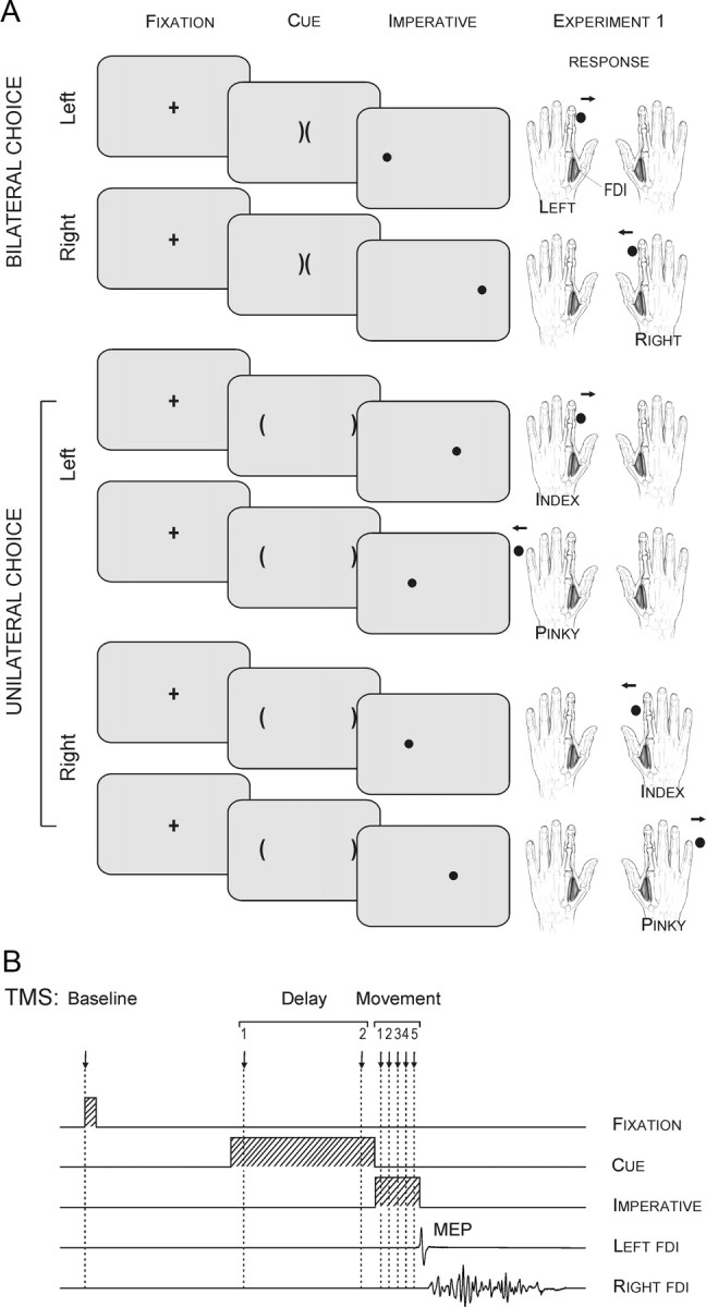
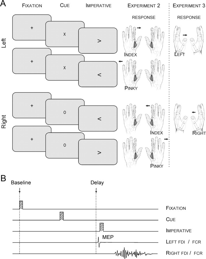
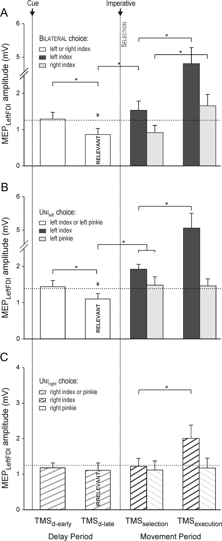
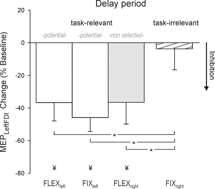
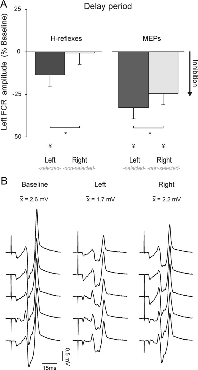
Inhibitory mechanisms are critically involved in goal-directed behaviors. To gain further insight into how such mechanisms shape motor representations during response preparation, motor evoked potentials (MEPs) elicited by transcranial magnetic stimulation (TMS) and H-reflexes were recorded from left hand muscles during choice reaction time tasks. The imperative signal, which indicated the required response, was always preceded by a preparatory cue. During the postcue delay period, left MEPs were suppressed when the left hand had been cued for the forthcoming response, suggestive of a form of inhibition specifically directed at selected response representations. H-reflexes were also suppressed on these trials, indicating that the effects of this inhibition extend to spinal circuits. In addition, left MEPs were suppressed when the right hand was cued, but only when left hand movements were a possible response option before the onset of the cue. Notably, left hand H-reflexes were not modulated on these trials, consistent with a cortical locus of inhibition that lowers the activation of task-relevant, but nonselected responses. These results suggest the concurrent operation of two inhibitory mechanisms during response preparation: one decreases the activation of selected responses at the spinal level, helping to control when selected movements should be initiated by preventing their premature release; a second, upstream mechanism helps to determine what response to make during a competitive selection process.


Figures

Figure 1.
Figure 1.
A, The preparatory cue consisted of two brackets, the “soccer goals.” The imperative, a circle or “soccer ball,” indicated the required response in all tasks. Participants were asked to shoot the ball in the appropriate goal by making an abduction movement of the corresponding index finger or pinky. Note that in the unileft and uniright tasks, subjects were asked to imagine they had the left or right hand, respectively, in the middle of the screen; in the bilateral task, the hands were imagined on the sides of the screen. B, Sequence and timing of events in experiment 1: A fixation marker (100 ms) was followed 900 ms later by a preparatory cue that lasted for a fixed delay (900 ms). An imperative signal then appeared (300 ms), indicating that the response should be initiated. A single TMS pulse was applied over the right M1 at eight possible timings during one of three epochs (baseline, delay, movement).
Figure 2.
Figure 2.
A, The preparatory cue consisted of a letter: “x” indicated a forthcoming left hand movement and “o” indicated a right hand movement. The imperative signal was an arrow presented at the screen center. B, Sequence and timing of events in experiments 2 and 3: A fixation marker (100 ms) was followed 900 ms later, by a preparatory cue that lasted for a fixed delay (100 ms). An imperative signal indicating the required response appeared after a delay (Exp. 2: 900 ms; Exp. 3: 900–1200 ms). A single TMS or electrical pulse was applied over the right M1 or the left median nerve, respectively, during one of two epochs (baseline, delay). In this example, a TMS pulse was applied during the delay period of a right index finger abduction (Exp. 2) or wrist flexion (Exp. 3) movement.
Figure 3.
Figure 3.
Amplitude (in millivolts) of MEPs recorded from left FDI following right M1 TMS in the bilateral (A), unileft (B), and uniright (C) tasks in experiment 1. MEP amplitudes are shown for the delay (following cue) and movement (following imperative) periods. The horizontal dashed line represents the baseline MEP amplitude. Left FDI MEP amplitudes were lower at the end of the delay period (TMSd-late) when the index finger was a potential respondent (bilateral and unileft) but not when it was task irrelevant (uniright). MEP amplitudes increased when the imperative signaled a left index finger response (TMSselection and TMSexecution). *p < 0.05. ¥Significant difference between MEPs at TMSd-late and TMSbaseline.
Figure 4.
Figure 4.
Percentage change, relative to baseline, in the amplitude of left FDI MEPs in experiment 2, recorded just before the imperative signal (TMSdelay). The “fixed” condition defines either left or right hand trials only. The “flexible” condition mixes left and right hand trials. Left FDI MEP suppression was evident when the left index finger was a potential respondent (fixleft, flexleft) and after a right hand cue in the flexible condition (flexright).
Figure 5.
Figure 5.
A, Percentage change in H-reflex (left histograms, n = 11) or MEP (right histograms, n = 11) amplitude recorded from the left FCR (flexor carpi radialis) at the end of the delay period. Data are expressed as a function of baseline values. *p < 0.05. ¥Significant difference with respect to baseline. B, Individual H-reflexes recorded from the left FCR in experiment 3 from a representative subject during the baseline epoch or delay period following a left or right hand cue.

Similar articles

Cited by

References

    1. Aron AR. The neural basis of inhibition in cognitive control. Neuroscientist. 2007;13:214–228. - PubMed
    1. Aron AR, Robbins TW, Poldrack RA. Inhibition and the right inferior frontal cortex. Trends Cogn Sci. 2004;8:170–177. - PubMed
    1. Bastian A, Schöner G, Riehle A. Preshaping and continuous evolution of motor cortical representations during movement preparation. Eur J Neurosci. 2003;18:2047–2058. - PubMed
    1. Beurze SM, de Lange FP, Toni I, Medendorp WP. Integration of target and effector information in the human brain during reach planning. J Neurophysiol. 2007;97:188–199. - PubMed
    1. Botvinick MM, Braver TS, Barch DM, Carter CS, Cohen JD. Conflict monitoring and cognitive control. Psychol Rev. 2001;108:624–652. - PubMed

Publication types

LinkOut - more resources