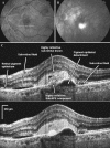
High-resolution Fourier-domain optical coherence tomography of choroidal neovascular membranes associated with age-related macular degeneration
- PMID: 20220054
- PMCID: PMC2910645
- DOI: 10.1167/iovs.09-4256
High-resolution Fourier-domain optical coherence tomography of choroidal neovascular membranes associated with age-related macular degeneration
Abstract
Purpose: To investigate the use of high-resolution Fourier-domain optical coherence tomography (Fd-OCT) to image choroidal neovascular membranes (CNVMs) associated with exudative age-related macular degeneration (eAMD).
Methods: An Fd-OCT instrument with axial resolution of 4 to 4.5 microm and transverse resolution of 10 to 15 microm was used to image 21 eyes (19 subjects) with newly diagnosed eAMD. A raster series of 100 B-scans separated by 60 microm was used to study the growth pattern of CNVM and associated morphologic changes. CNVM size was determined using 250 to 300 serial virtual C-scans of reconstructed three-dimensional macular volume.
Results: A highly reflective subretinal and/or subretinal pigment epithelial (RPE) lesion that co-localized with the CNVM seen on fluorescein angiography was detected in all eyes by Fd-OCT. Although a combined subretinal and sub-RPE growth pattern of various degrees was noted in 15 (71%) eyes, a statistically significant difference in the distribution of growth pattern was noted when classic CNVM was compared with occult CNVM (chi(2) = 10.4, df = 2, P < 0.005). Classic lesions had >90% subretinal growth pattern, whereas occult lesions had a more variable growth pattern. Angiographic CNVM size correlated with size on Fd-OCT but correlation was better for classic CNVM (classic, r = 0.99, P < 0.0001; nonclassic, r = 0.78, P < 0.001).
Conclusions: Fd-OCT is a promising potential alternative modality to visualize CNVM with AMD. Angiographic lesion size and type correlated with growth pattern and size of CNVM on Fd-OCT, with correlation being stronger for classic lesions.
Figures


References
-
- Friedman DS, O'Colmain BJ, Munoz B, et al. for the Eye Diseases Prevalence Research Group. Prevalence of age-related macular degeneration in the United States. Arch Ophthalmol 2004;122:564–572 - PubMed
-
- Green WR, McDonnell PJ, Yeo JH. Pathologic features of senile macular degeneration. Ophthalmology 1985;92:615–627 - PubMed
-
- Green WR. Clinicopathologic studies of treated choroidal neovascular membranes: a review and report of two cases. Retina 1991;11:328–356 - PubMed
-
- Green WR, Enger C. Age-related macular degeneration histopathologic studies. Ophthalmology 1993;100:1519–1535 - PubMed
-
- Grossniklaus HE, Hutchinson AK, Capone A, Jr, et al. Clinicopathologic features of surgically-excised choroidal neovascular membranes. Ophthalmology 1994;101:1099–1111 - PubMed
Publication types
MeSH terms
Grants and funding
LinkOut - more resources
Full Text Sources
Medical
Miscellaneous

