

Intravitreous delivery of the corticosteroid fluocinolone acetonide attenuates retinal degeneration in S334ter-4 rats
- PMID: 20220055
- PMCID: PMC2910647
- DOI: 10.1167/iovs.09-4492
Intravitreous delivery of the corticosteroid fluocinolone acetonide attenuates retinal degeneration in S334ter-4 rats
Abstract
Purpose: To study the neuroprotective properties of low-dose, sustained-release intravitreous fluocinolone acetonide (FA) in transgenic S334ter-4 rats.
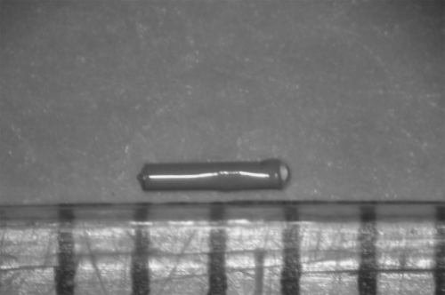
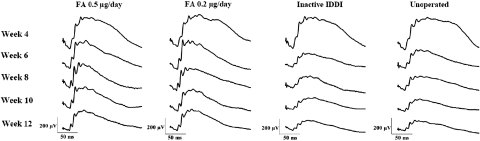
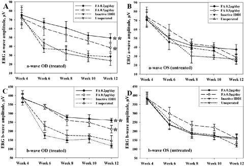
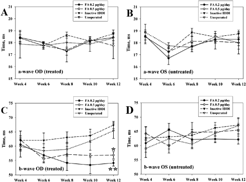
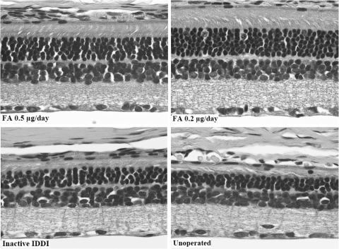
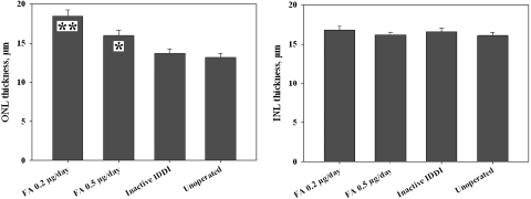
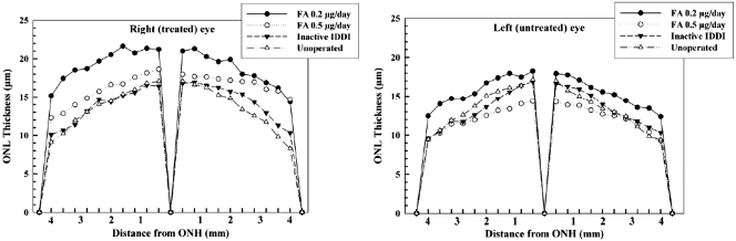
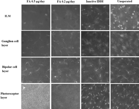
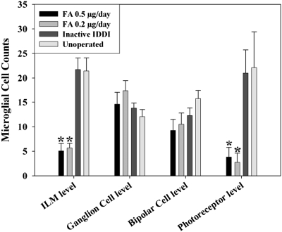
Methods: S334ter-4 rats aged 4 weeks were divided into four groups: 0.5 microg/d FA-loaded intravitreous drug delivery implant (IDDI); 0.2 microg/d FA-loaded IDDI; inactive IDDI; and unoperated controls. Electroretinography (ERG) was performed before surgery and every 2 weeks after surgery for 8 weeks. When the rats were 12 weeks of age, outer nuclear layer (ONL) and inner nuclear layer (INL) thicknesses were measured. Microglial cell counts were obtained from retinal wholemounts labeled for Iba-1.
Results: At the end of the study, unoperated and inactive IDDI-implanted rats demonstrated 50% to 60% reductions in ERG amplitudes compared with those recorded at 4 weeks (P < 0.001 for both groups). FA 0.2-microg/d animals demonstrated 15% amplitude attenuation, while FA 0.5-microg/d animals showed 30% reduction. ONL thickness in FA 0.2-microg/d-treated eyes was 25.8% +/- 2.3% higher than in control group eyes (P < 0.001) and 30.0% +/- 2.1% higher than in inactive IDDI-implanted eyes (P < 0.001). In FA 0.5-microg/d-treated eyes, ONL thickness was 22.4% +/- 2.8% higher than in control group eyes (P < 0.001) and 22.3% +/- 3.7% higher than in inactive IDDI-implanted eyes (P < 0.01). No statistically significant difference was observed between the two control groups. No statistically significant difference between the two FA-treated groups was found. FA-treated groups demonstrated significantly fewer activated microglial cells than control groups.
Conclusions: Chronic intravitreous infusion of FA preserves ONL cell morphology and ERG a- and b-wave amplitudes and reduces retinal neuroinflammation in S334ter rats. Based on these findings, the synthetic corticosteroid FA may promise a therapeutic role in patients with retinal degeneration.
Figures
Similar articles
-
Photoreceptor neuroprotection in RCS rats via low-dose intravitreal sustained-delivery of fluocinolone acetonide.Invest Ophthalmol Vis Sci. 2009 Oct;50(10):4847-57. doi: 10.1167/iovs.08-2831. Epub 2009 Apr 30. Invest Ophthalmol Vis Sci. 2009. PMID: 19407016
-
Neuroprotective effect of subretinal implants in the RCS rat.Invest Ophthalmol Vis Sci. 2005 Feb;46(2):674-82. doi: 10.1167/iovs.04-0515. Invest Ophthalmol Vis Sci. 2005. PMID: 15671299
-
Safety and pharmacokinetics of an intraocular fluocinolone acetonide sustained delivery device.Invest Ophthalmol Vis Sci. 2000 Oct;41(11):3569-75. Invest Ophthalmol Vis Sci. 2000. PMID: 11006254
-
Influence of eye pigmentation on retinal degeneration in P23H and S334ter mutant rhodopsin transgenic rats.Exp Eye Res. 2019 Oct;187:107755. doi: 10.1016/j.exer.2019.107755. Epub 2019 Aug 10. Exp Eye Res. 2019. PMID: 31408630
-
Sustained-release fluocinolone acetonide intravitreal insert for macular edema: clinical pharmacology and safety evaluation.Expert Opin Drug Saf. 2015 Jul;14(7):1147-56. doi: 10.1517/14740338.2015.1041916. Epub 2015 May 20. Expert Opin Drug Saf. 2015. PMID: 25994877 Review.
Cited by
-
Long-term photoreceptor rescue in two rodent models of retinitis pigmentosa by adeno-associated virus delivery of Stanniocalcin-1.Exp Eye Res. 2017 Dec;165:175-181. doi: 10.1016/j.exer.2017.09.011. Epub 2017 Sep 30. Exp Eye Res. 2017. PMID: 28974356 Free PMC article.
-
Therapeutic Options in Refractory Diabetic Macular Oedema.Drugs. 2017 Apr;77(5):481-492. doi: 10.1007/s40265-017-0704-6. Drugs. 2017. PMID: 28197794 Review.
-
Chronic stress and corticosterone exacerbate alcohol-induced tissue injury in the gut-liver-brain axis.Sci Rep. 2021 Jan 12;11(1):826. doi: 10.1038/s41598-020-80637-y. Sci Rep. 2021. PMID: 33436875 Free PMC article.
-
The role of microglia in diabetic retinopathy.J Ophthalmol. 2014;2014:705783. doi: 10.1155/2014/705783. Epub 2014 Aug 31. J Ophthalmol. 2014. PMID: 25258680 Free PMC article. Review.
-
Dendrimers as tunable vectors of drug delivery systems and biomedical and ocular applications.Int J Nanomedicine. 2015 Dec 22;11:1-12. doi: 10.2147/IJN.S93069. eCollection 2016. Int J Nanomedicine. 2015. PMID: 26730187 Free PMC article. Review.
References
-
- Kreutzberg GW. Microglia: a sensor for pathological events in the CNS. Trends Neurosci 1996;19:312–318 - PubMed
-
- Kreutzberg GW. Microglia, the first line of defence in brain pathologies. Arzneimittel-Forschung 1995;45:357–360 - PubMed
-
- Nakanishi H. Microglial functions and proteases. Mol Neurobiol 2003;27:163–176 - PubMed
-
- Nichols NR. Glial responses to steroids as markers of brain aging. J Neurobiol 1999;40:585–601 - PubMed
Publication types
MeSH terms
Substances
Grants and funding
LinkOut - more resources
Full Text Sources
Other Literature Sources
Medical
