Role of matrix metalloproteinase-9 in the development of diabetic retinopathy and its regulation by H-Ras
- PMID: 20220057
- PMCID: PMC2910650
- DOI: 10.1167/iovs.09-4851
Role of matrix metalloproteinase-9 in the development of diabetic retinopathy and its regulation by H-Ras
Abstract
Purpose: Diabetes activates a small molecular weight G-protein, H-Ras, in the retina and its capillary cells, and H-Ras activation is implicated in the apoptosis of retinal capillary cells. Matrix metalloproteinase (MMP)-9 is regulated by H-Ras, and in diabetes its activation is associated with increased vascular permeability. The goal of this study was to investigate the role of sustained activation of MMP-9 in the pathogenesis of diabetic retinopathy and to illustrate the mechanism through which it is upregulated in diabetes.
Methods: Retinal MMP-9 activation and its tissue inhibitor, TIMP-1, were quantified in streptozotocin-induced diabetic rats. Inhibition of H-Ras by simvastatin on diabetes-induced activation of H-Ras was evaluated. The mechanism by which diabetes regulates retinal MMP-9 was confirmed by determining the effect of genetic or pharmacologic regulation of H-Ras on its activation in retinal endothelial cells.
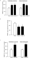
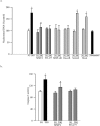
Results: In rats, MMP-9 was activated and expression of TIMP-1 was decreased in the retina and its microvasculature at both 2 months and 12 months of diabetes. In retinal endothelial cells, high glucose activated MMP-9, and inhibition of its activation (by pharmacologic inhibitor or siRNA) ameliorated accelerated apoptosis. Inhibition of H-Ras, both in diabetic rats (simvastatin) and in isolated endothelial cells (H-Ras siRNA), abrogated the activation of MMP-9 and prevented the reduction of TIMP-1.
Conclusions: Hyperglycemia-induced activation of MMP-9 accelerates apoptosis of retinal capillary cells, a phenomenon that predicts the development of diabetic retinopathy, and the activation of MMP-9 is downstream of H-Ras. Characterizing the role of MMP-9 in the development of diabetic retinopathy will help explore novel molecular targets for future pharmacological interventions.
Figures


Similar articles
-
Abrogation of MMP-9 gene protects against the development of retinopathy in diabetic mice by preventing mitochondrial damage.Diabetes. 2011 Nov;60(11):3023-33. doi: 10.2337/db11-0816. Epub 2011 Sep 20. Diabetes. 2011. PMID: 21933988 Free PMC article.
-
Molecular Mechanism of Transcriptional Regulation of Matrix Metalloproteinase-9 in Diabetic Retinopathy.J Cell Physiol. 2016 Aug;231(8):1709-18. doi: 10.1002/jcp.25268. Epub 2015 Dec 22. J Cell Physiol. 2016. PMID: 26599598
-
Oxidative stress and the development of diabetic retinopathy: contributory role of matrix metalloproteinase-2.Free Radic Biol Med. 2009 Jun 15;46(12):1677-85. doi: 10.1016/j.freeradbiomed.2009.03.024. Epub 2009 Apr 5. Free Radic Biol Med. 2009. PMID: 19345729 Free PMC article.
-
Matrix metalloproteinases in diabetic retinopathy: potential role of MMP-9.Expert Opin Investig Drugs. 2012 Jun;21(6):797-805. doi: 10.1517/13543784.2012.681043. Epub 2012 Apr 23. Expert Opin Investig Drugs. 2012. PMID: 22519597 Free PMC article. Review.
-
Regulation of Matrix Metalloproteinase in the Pathogenesis of Diabetic Retinopathy.Prog Mol Biol Transl Sci. 2017;148:67-85. doi: 10.1016/bs.pmbts.2017.02.004. Epub 2017 Mar 14. Prog Mol Biol Transl Sci. 2017. PMID: 28662829 Review.
Cited by
-
The α7-nicotinic acetylcholine receptor and MMP-2/-9 pathway mediate the proangiogenic effect of nicotine in human retinal endothelial cells.Invest Ophthalmol Vis Sci. 2011 Jun 22;52(7):4428-38. doi: 10.1167/iovs.10-5461. Invest Ophthalmol Vis Sci. 2011. PMID: 20554619 Free PMC article.
-
Hyperglycemia induces altered expressions of angiogenesis associated molecules in the trophoblast.Evid Based Complement Alternat Med. 2013;2013:457971. doi: 10.1155/2013/457971. Epub 2013 Jul 25. Evid Based Complement Alternat Med. 2013. PMID: 23983782 Free PMC article.
-
Statins for prevention of diabetic-related blindness: a new treatment option?Expert Rev Ophthalmol. 2011 Jun;6(3):269-272. doi: 10.1586/eop.11.36. Expert Rev Ophthalmol. 2011. PMID: 21938261 Free PMC article. No abstract available.
-
Abrogation of MMP-9 gene protects against the development of retinopathy in diabetic mice by preventing mitochondrial damage.Diabetes. 2011 Nov;60(11):3023-33. doi: 10.2337/db11-0816. Epub 2011 Sep 20. Diabetes. 2011. PMID: 21933988 Free PMC article.
-
Proteomic profiling reveals immunomodulatory role of IL-33 in ocular bacterial and fungal infections.Infect Immun. 2025 Jul 8;93(7):e0018325. doi: 10.1128/iai.00183-25. Epub 2025 Jun 13. Infect Immun. 2025. PMID: 40512033 Free PMC article.
References
-
- Kern TS, Tang J, Mizutani M, Kowluru R, Nagraj R, Lorenzi M. Response of capillary cell death to aminoguanidine predicts the development of retinopathy: comparison of diabetes and galactosemia. Invest Ophthalmol Vis Sci 2000;41:3972–3978 - PubMed
-
- Kowluru RA, Abbas SN. Diabetes-induced mitochondrial dysfunction in the retina. Invest Ophthalmol Vis Sci 2003;44:5327–5334 - PubMed
Publication types
MeSH terms
Substances
Grants and funding
LinkOut - more resources
Full Text Sources
Other Literature Sources
Medical
Research Materials
Miscellaneous

