

Activity-dependent release of endogenous BDNF from mossy fibers evokes a TRPC3 current and Ca2+ elevations in CA3 pyramidal neurons
- PMID: 20220070
- PMCID: PMC2867575
- DOI: 10.1152/jn.01140.2009
Activity-dependent release of endogenous BDNF from mossy fibers evokes a TRPC3 current and Ca2+ elevations in CA3 pyramidal neurons
Abstract
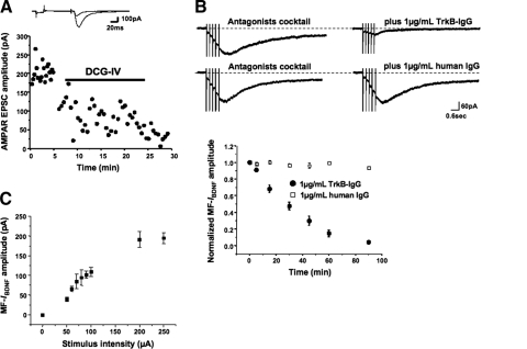
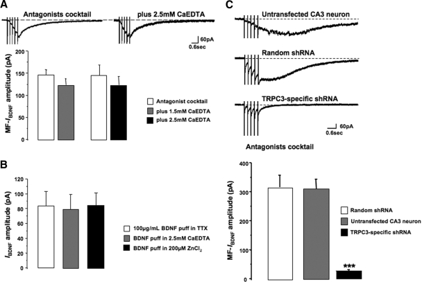
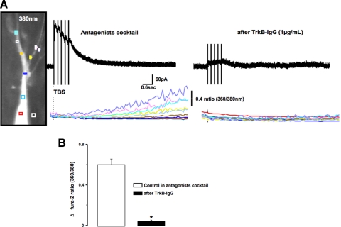
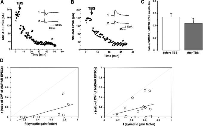
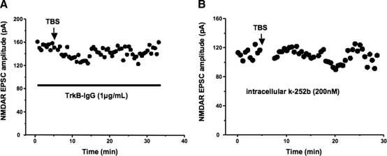
Multiple studies have demonstrated that brain-derived neurotrophic factor (BDNF) is a potent modulator of neuronal structure and function in the hippocampus. However, the majority of studies to date have relied on the application of recombinant BDNF. We herein report that endogenous BDNF, released via theta burst stimulation of mossy fibers (MF), elicits a slowly developing cationic current and intracellular Ca(2+) elevations in CA3 pyramidal neurons with the same pharmacological profile of the transient receptor potential canonical 3 (TRPC3)-mediated I(BDNF) activated in CA1 neurons by brief localized applications of recombinant BDNF. Indeed, sensitivity to both the extracellular BDNF scavenger tropomyosin-related kinase B (TrkB)-IgG and small hairpin interference RNA-mediated TRPC3 channel knockdown confirms the identity of this conductance as such, henceforth-denoted MF-I(BDNF). Consistent with such activity-dependent release of BDNF, these MF-I(BDNF) responses were insensitive to manipulations of extracellular Zn(2+) concentration. Brief theta burst stimulation of MFs induced a long-lasting depression in the amplitude of excitatory postsynaptic currents (EPSCs) mediated by both AMPA and N-methyl-d-aspartate (NMDA) receptors without changes in the NMDA receptor/AMPA receptor ratio, suggesting a reduction in neurotransmitter release. This depression of NMDAR-mediated EPSCs required activity-dependent release of endogenous BDNF from MFs and activation of Trk receptors, as it was sensitive to the extracellular BDNF scavenger TrkB-IgG and the tyrosine kinase inhibitor k-252b. These results uncovered the most immediate response to endogenously released--native--BDNF in hippocampal neurons and lend further credence to the relevance of BDNF signaling for synaptic function in the hippocampus.
Figures
Similar articles
-
TRPC3 channels are necessary for brain-derived neurotrophic factor to activate a nonselective cationic current and to induce dendritic spine formation.J Neurosci. 2007 May 9;27(19):5179-89. doi: 10.1523/JNEUROSCI.5499-06.2007. J Neurosci. 2007. PMID: 17494704 Free PMC article.
-
Depolarization-induced long-term depression at hippocampal mossy fiber-CA3 pyramidal neuron synapses.J Neurosci. 2003 Oct 29;23(30):9786-95. doi: 10.1523/JNEUROSCI.23-30-09786.2003. J Neurosci. 2003. PMID: 14586006 Free PMC article.
-
LTD at mossy fiber synapses onto stratum lucidum interneurons requires TrkB and retrograde endocannabinoid signaling.J Neurophysiol. 2019 Feb 1;121(2):609-619. doi: 10.1152/jn.00669.2018. Epub 2018 Dec 5. J Neurophysiol. 2019. PMID: 30517040 Free PMC article.
-
Role of giant depolarizing potentials in shaping synaptic currents in the developing hippocampus.Crit Rev Neurobiol. 2006;18(1-2):13-23. doi: 10.1615/critrevneurobiol.v18.i1-2.30. Crit Rev Neurobiol. 2006. PMID: 17725505 Review.
-
BDNF and Lactate as Modulators of Hippocampal CA3 Network Physiology.Cell Mol Neurobiol. 2023 Nov;43(8):4007-4022. doi: 10.1007/s10571-023-01425-6. Epub 2023 Oct 24. Cell Mol Neurobiol. 2023. PMID: 37874456 Free PMC article. Review.
Cited by
-
Contribution of TRPC Channels in Neuronal Excitotoxicity Associated With Neurodegenerative Disease and Ischemic Stroke.Front Cell Dev Biol. 2021 Jan 8;8:618663. doi: 10.3389/fcell.2020.618663. eCollection 2020. Front Cell Dev Biol. 2021. PMID: 33490083 Free PMC article.
-
The physiology of regulated BDNF release.Cell Tissue Res. 2020 Oct;382(1):15-45. doi: 10.1007/s00441-020-03253-2. Epub 2020 Sep 18. Cell Tissue Res. 2020. PMID: 32944867 Free PMC article. Review.
-
Canonical Transient Receptor Potential Channel 3 Contributes to Febrile Seizure Inducing Neuronal Cell Death and Neuroinflammation.Cell Mol Neurobiol. 2018 Aug;38(6):1215-1226. doi: 10.1007/s10571-018-0586-5. Epub 2018 May 10. Cell Mol Neurobiol. 2018. PMID: 29748835 Free PMC article.
-
Discovery of a Highly Selective and Potent TRPC3 Inhibitor with High Metabolic Stability and Low Toxicity.ACS Med Chem Lett. 2021 Mar 5;12(4):572-578. doi: 10.1021/acsmedchemlett.0c00571. eCollection 2021 Apr 8. ACS Med Chem Lett. 2021. PMID: 33859797 Free PMC article.
-
Pharmacologically targeting transient receptor potential channels for seizures and epilepsy: Emerging preclinical evidence of druggability.Pharmacol Ther. 2023 Apr;244:108384. doi: 10.1016/j.pharmthera.2023.108384. Epub 2023 Mar 16. Pharmacol Ther. 2023. PMID: 36933703 Free PMC article. Review.
References
Publication types
MeSH terms
Substances
Grants and funding
LinkOut - more resources
Full Text Sources
Miscellaneous