. 2010 May;103(5):2570-80.
doi: 10.1152/jn.00013.2010. Epub 2010 Mar 10.

Impaired glial glutamate uptake induces extrasynaptic glutamate spillover in the spinal sensory synapses of neuropathic rats

Affiliations

Impaired glial glutamate uptake induces extrasynaptic glutamate spillover in the spinal sensory synapses of neuropathic rats

Hui Nie et al. J Neurophysiol. 2010 May.

Abstract

Glial cell dysfunction and excessive glutamate receptor activation in spinal dorsal horn neurons are hallmark mechanisms of pathological pain. The way in which glial cell dysfunction leads to excessive glutamate receptor activation in the spinal sensory synapses remains unknown. We and others recently reported the downregulation of glial glutamate transporter (GT) protein expression in the spinal dorsal horn of neuropathic rats. In this study, we showed that excitatory postsynaptic currents originating from N-methyl-d-aspartate receptor activation (NMDA EPSCs) elicited by peripheral synaptic input in the spinal sensory synapses were enhanced in neuropathic rats with mechanical allodynia induced by partial sciatic nerve ligation. The enhanced NMDA EPSCs were accompanied by an increased proportion of NR2B receptor activation. Physically blocking the extrasynaptic glutamate with dextran or chemically scavenging the glutamate with glutamic-pyruvic transaminase ameliorated the abnormal NMDA EPSCs in neuropathic rats. Pharmacological blockade of glial GTs with dihydrokainic acid enhanced NMDA receptor activation elicited by synaptic input or puffed glutamate in normal control rats, but this effect was precluded in neuropathic rats. Thus extrasynaptic glutamate spillover and extrasynaptic NMDA receptor activation induced by deficient glial glutamate uptake in the synapses resulted in the excessive activation of NMDA receptors in neuropathic rats. It is suggested that extrasynaptic glutamate spillover may be a key synaptic mechanism related to phenotypic alterations induced by nerve injury in the spinal dorsal horn and that glial GTs are potential new targets in the development of analgesics.


Figures

Fig. 1.
Fig. 1.
Enhanced activation of N-methyl-d-aspartate (NMDA) receptors in spinal superficial dorsal horn neurons in neuropathic rats. A: samples of NMDA excitatory postsynaptic currents (NMDA EPSCs) evoked by 2T and maximum stimulation in control and neuropathic rats. B: bar graphs showing the mean ± SE amplitudes, peak latencies, durations, decay time constants, and areas of EPSCs. *Comparisons of EPSCs in control and neuropathic rats. **P < 0.01; ***P < 0.001.
Fig. 2.
Fig. 2.
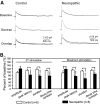
Spinal superficial dorsal horn neurons in neuropathic rats had an increased proportion of NR2B receptors activated by peripheral input. A: original recordings showing samples of NMDA EPSCs evoked by 2T and maximum stimulation at baseline and in the presence of a NR2B receptor-specific antagonist (Ro 25–6981, 1 μM) in control rats (left) and neuropathic rats (right). B: bar graphs showing the mean amplitudes, peak latencies, durations, and decay time constants of the NMDA EPSCs evoked by 2T and maximum stimulation in the presence of Ro 25–6981 in control and neuropathic rats. All values were normalized to the baseline values prior to application of Ro 25–6981. *Comparison of the effects induced by Ro 25–6981 on NMDA EPSCs in control and neuropathic rats. *P < 0.05; **P < 0.01.
Fig. 3.
Fig. 3.
Limiting glutamate spillover with 5% dextran reduced the NMDA EPSC peak latency and duration and increased the NMDA EPSC peak amplitude in neuropathic rats but did not alter the NMDA EPSC in control rats. A: original recordings showing samples of NMDA EPSCs evoked by maximum stimulation in spinal superficial dorsal horn neurons in control (left) and neuropathic (right) rats recorded before (top) and during (middle) perfusion of 5% dextran. Bottom: overlaps of the top and middle. B: bar graphs showing the mean percentage changes in the NMDA EPSC amplitude, peak latency, duration, and decay time constant induced by dextran. *Comparisons of the effects induced by dextran on NMDA EPSCs in control and neuropathic rats. *P < 0.05; ***P < 0.001.
Fig. 4.
Fig. 4.
Limiting extrasynaptic glutamate spillover with the glutamate scavenger glutamic-pyruvic transaminase reduced the NMDA EPSC peak amplitude, latency, duration, and decay time constant in neuropathic rats but not in control rats. A: original recordings showing samples of NMDA EPSCs evoked by maximum stimulation in spinal superficial dorsal horn neurons in control (left) and neuropathic (right) rats recorded before (top) and during perfusion (middle), and after washout (bottom) of the glutamate scavenger. B: bar graphs showing the mean percentage changes in the NMDA EPSC amplitude, peak latency, duration, and decay time constant induced by the scavenger. *Comparison of the effects induced by the scavenger on NMDA EPSCs between control and neuropathic rats. *P < 0.05; **P < 0.01; ***P < 0.001.
Fig. 5.
Fig. 5.
Impairment of glutamate clearance by glial glutamate transporters (GTs) in neuropathic rats is key to prolonged activation of NMDA receptors evoked by exogenous glutamate in neuropathic rats. A: original recordings showing NMDA currents evoked by puff application (20 ms) of l-glutamate (glu-NMDA current) and NMDA (agonist-NMDA current) recorded, respectively, in the same neuron at baseline and during perfusion of dihydrokainic acid (DHK, 300 μM) in control and neuropathic rats. ↑, onset of puff application. The recordings were obtained for spinal superficial dorsal horn neurons in the presence of TTX (1 μM) and 6,7-dinitroquinoxaline-2,3-dione (DNQX, 10 μM) at a holding potential of +40 mV. B: mean amplitudes, peak latencies, durations, and decay time constants for glu-NMDA current and agonist-NMDA current in control rats and neuropathic rats. C: mean percentage changes induced by DHK in glu-NMDA current and agonist-NMDA current in control and neuropathic rats. *Comparison between control and neuropathic rats. *P < 0.05. **P < 0.01. ***P < 0.001.
Fig. 6.
Fig. 6.
Impairment of glutamate uptake by glial GTs causes prolonged activation of NMDA receptors in spinal sensory synapses activated by peripheral input in neuropathic rats. A: original recordings showing samples of NMDA EPSCs evoked by 2T and maximum stimulation in spinal superficial dorsal horn neurons in control and neuropathic rats obtained before (baseline) and during perfusion of DHK (300 μM). B: bar graphs showing the mean percentages of changes in the NMDA EPSC amplitude, peak latency, duration, and decay time constant induced by DHK. *Comparison of the effects on NMDA EPSCs induced by DHK in control and neuropathic rats. *P < 0.05. **P < 0.01.

References

    1. Agulhon C, Petravicz J, McMullen AB, Sweger EJ, Minton SK, Taves SR, Casper KB, Fiacco TA, McCarthy KD. What is the role of astrocyte calcium in neurophysiology? Neuron 59: 932–946, 2008. - PMC - PubMed
    1. Anderson CM, Swanson RA. Astrocyte glutamate transport: review of properties, regulation, and physiological functions. GLIA 32: 1–14, 2000. - PubMed
    1. Angulo MC, Kozlov AS, Charpak S, Audinat E. Glutamate released from glial cells synchronizes neuronal activity in the hippocampus. J Neurosci 24: 6920–6927, 2004. - PMC - PubMed
    1. Arriza JL, Fairman WA, Wadiche JI, Murdoch GH, Kavanaugh MP, Amara SG. Functional comparisons of three glutamate transporter subtypes cloned from human motor cortex. J Neurosci 14: 5559–5569, 1994. - PMC - PubMed
    1. Baba H, Doubell TP, Moore KA, Woolf CJ. Silent NMDA receptor-mediated synapses are developmentally regulated in the dorsal horn of the rat spinal cord. J Neurophysiol 83: 955–962, 2000. - PubMed

Publication types

MeSH terms

LinkOut - more resources