. 2010 Apr 15;184(8):4440-6.
doi: 10.4049/jimmunol.0902984. Epub 2010 Mar 10.

Plasmacytoid dendritic cells enhance mortality during lethal influenza infections by eliminating virus-specific CD8 T cells

Affiliations

Plasmacytoid dendritic cells enhance mortality during lethal influenza infections by eliminating virus-specific CD8 T cells

Ryan A Langlois et al. J Immunol. .

Abstract

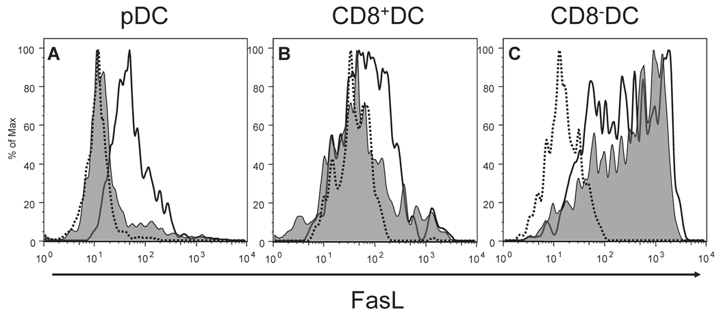
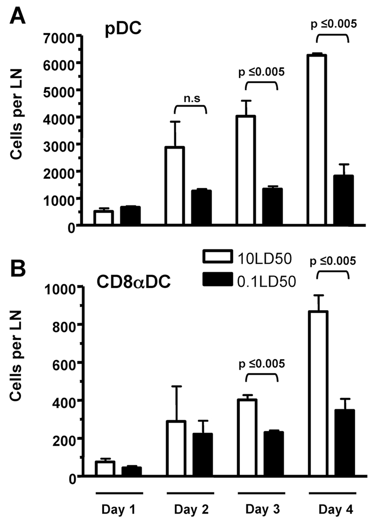
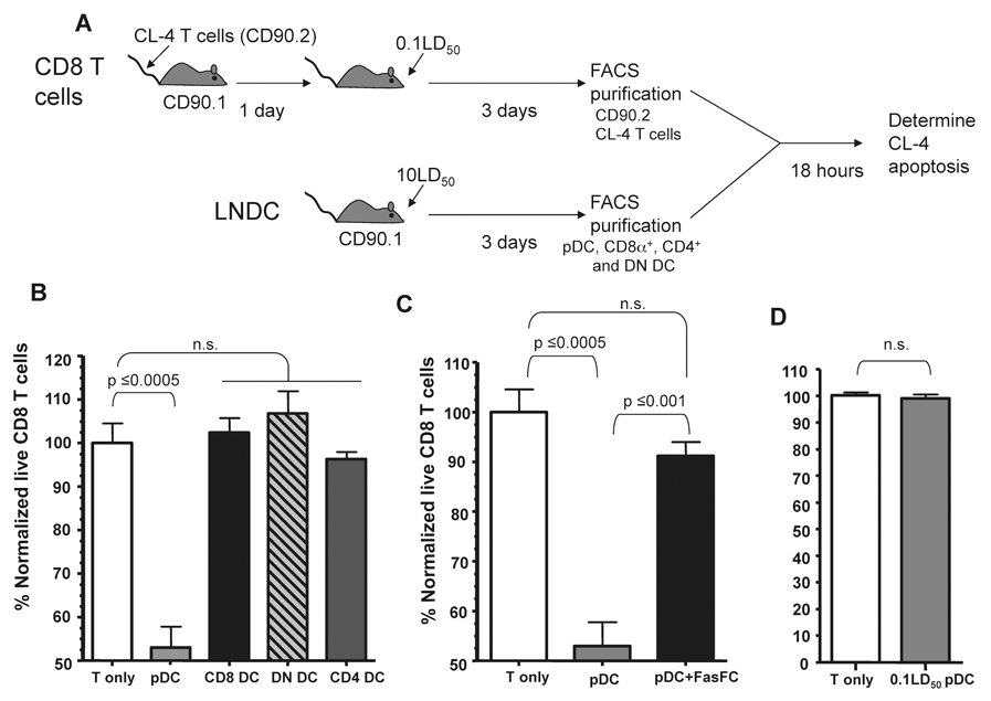
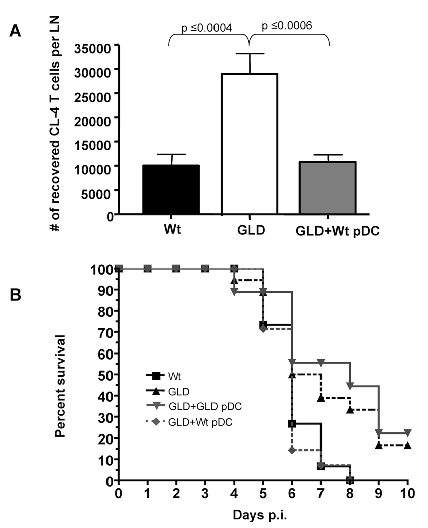
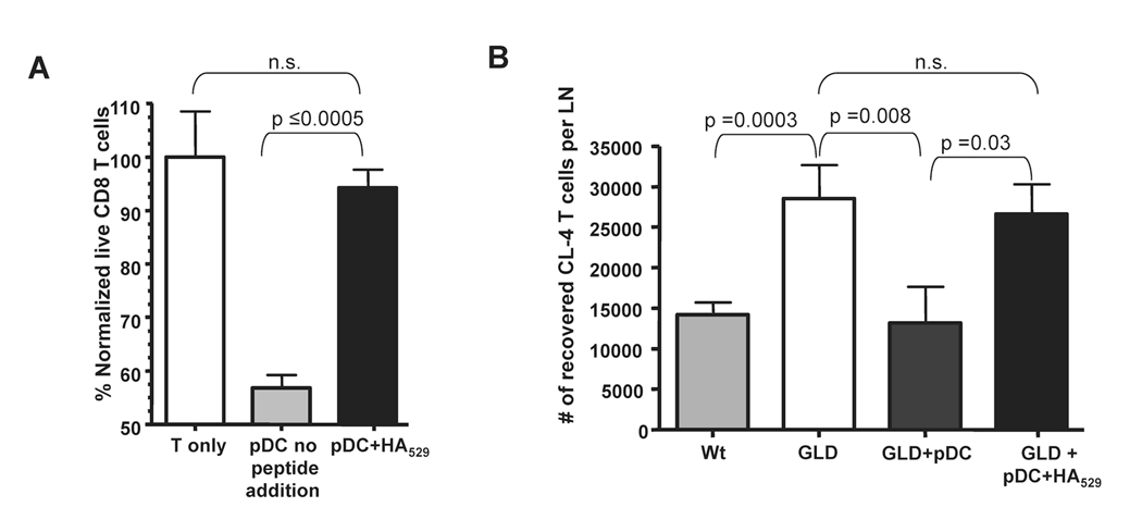
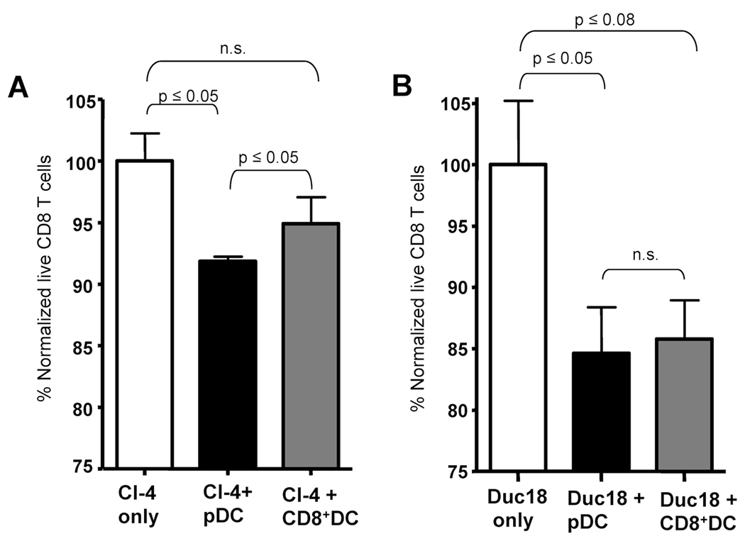
Previous studies have shown that the reduction in CD8 T cell immunity observed during high-dose influenza A virus (IAV) infection is mediated via lymph node (LN) dendritic cells (DCs) that express Fas ligand (FasL) and drive FasL-Fas (DC-T)-induced apoptosis. However, the specific DC subset(s) within the LN and the additional factors required for DC-mediated elimination of IAV-specific CD8 T cells remain unknown. In this paper, we demonstrate that plasmacytoid DCs (pDCs), which downregulate FasL during sublethal, but not lethal, IAV infection, accumulate to greater numbers within the LNs of lethal dose-infected mice. Further our findings show that pDCs from lethal, but not sublethal, dose IAV infections drive elimination of Fas(+) CD8 T cells and that this elimination occurs only in the absence of TCR recognition of IAV peptide-MHC class I complexes. Together, these results suggest that pDCs play a heretofore unknown deleterious role during lethal dose IAV infections by limiting the CD8 T cell response.


Figures

Figure 1
Figure 1
pDC and CD8α+ DC modulate FasL expression during IAV infection. Mice were infected with either a 10LD50 (black line) or 0.1LD50 (shaded histogram) of IAV and on day 3 p.i. cells from draining LN (pooled) from each group examined for FasL expression. Dotted line represents staining with an isotype control mAb. (A) FasL expression on CD11cmodCD45R+CD8+ cells (i.e. pDC); 10LD50 M.F.I.=60.5 and 0.1LD50 M.F.I.=28.3. (B) FasL expression on CD11c+CD45RCD8+ cells (i.e. CD8α+ DC); 10LD50 M.F.I.=139 and 0.1LD50 M.F.I.=118. (C) FasL expression on CD11c+CD45RCD8 cells (i.e. CD8α DC); 10LD50 M.F.I.=618 and 0.1LD50 M.F.I.=496.2. The MFI of staining with isotype control mAb has been subtracted from the FasL MFI for the DC subsets to yield the MFI reported above. Data are representative of 5 independent experiments.
Figure 2
Figure 2
pDC preferentially accumulate in the lung draining LN of lethal dose IAV infected mice. Mice were infected with IAV as in Fig. 1 and on day 1-4 p.i. LN from each group were pooled and the number of (A) CD11cmodCD45R+ cells (i.e. pDC) and (B) CD11c+CD45RCD8+ cells (i.e. CD8α+ DC) in the LN was determined. Data are representative of 3 independent experiments with 2–3 mice per group.
Figure 3
Figure 3
pDC from lethal dose IAV infected mice kill activated IAV-specific CD8 T cells. (A) Experimental setup for LNDC:CD8 T cell ex vivo apoptosis assay. (B) 104 of the indicated purified LNDC subsets were incubated with 104 activated CL-4 T cells and incubated at 37°C for 18 hours. After incubation the percentage of CD90.2+Annexin V7-AAD live CL-4 T cells was determined and normalized to CD8 T cells incubated alone (~70% live). Data are representative of 3 independent experiments. (C) pDC and CL-4 T cells were purified and incubated +/− Fas-Fc and the percentage of live CD8 T cells was determined as described above. Data are representative of 2 independent experiments. (D) 104 pDC from 0.1LD50 IAV-infected mice were incubated with 104 activated CL-4 T cells and apoptosis measured as in B. Data are representative of 3 independent experiments. n.s. = not significant.
Figure 4
Figure 4
Donnor wild-type pDC reduce IAV-specific T cell numbers and cause enhanced mortality in mice deficient in functional FasL. (A) CD90.2 wild type (Wt) and gld mice received 2×10⁶ CD90.1 CL-4 T cells and 24 hours post transfer were infected with a 10LD50 dose of IAV. 24 hours p.i. one group of gld mice received 2×10⁶ Wt pDC. On day 4 p.i. lung draining LN were removed and CD3+CD8+CD90.1+ Cl-4 T cells enumerated. Data are pooled from 2 independent experiments with 8–10 mice per group. (B) gld or wild type mice were infected with a 2.5LD50 of IAV. 24 hours p.i. pDC were purified from naïve wild-type or gld mice and then transferred (×106) into IAV-infected gld mice. Mortality was then monitored for 10 days p.i.. p values are as follows: Wt vs GLD=0.0285, Wt vs (GLD+ Wt pDC) =0.7013, GLD vs. (GLD+Wt pDC) =0.0157, (GLD+ Wt pDC) vs (GLD+ GLD pDC) =0.0103 and (GLD+ GLD pDC) vs GLD=0.5722. Data are pooled from 3 independent experiments with n values equaling; Wt=15, GLD+Wt pDC =14, GLD+ GLD pDC=9, GLD=18.
Figure 5
Figure 5
pDC induction of apoptosis in IAV-specific CD8 T cells is abrogated by cognate viral peptide:MHC I presentation. (A) CL-4 T cells and pDC were purified as described in Fig. 4, co-cultured +/− 1mM HA529 peptide and then the percentage of live CD8 T cells determined as in Fig. 4. (B) pDC were purified as in Fig. 4 and pulsed with 1µM HA529 peptide and transferred into IAV-infected mice and CL-4 T cell numbers measured as in Fig. 4. Data are representative of 2–3 independent experiments. n.s. = not significant.
Figure 6
Figure 6
pDC from lethal dose IAV infected mice eliminate IAV-specific and non IAV-specific activated CD8 T cells. CD8 T cells from the spleens of naïve CL-4 (A) and DUC18 (B) transgenic mice were purified and cultured for 3 days in the presence of αCD3/CD28 antibodies in order to activate the T cells. Activated transgenic T cells (104) were incubated with pDC or CD8α+DC (104) from lethal dose IAV-infected mice at 37°C for 18 hours as described in Figure 3. After incubation the percentage of CD90.2+Annexin V7-AAD live transgenic T cells was determined and normalized to CD8 T cells incubated alone. n.s.= p > 0.1. Data are representative of 2 independent experiments.

Similar articles

Cited by

References

    1. Eichelberger M, Allan W, Zijlstra M, Jaenisch R, Doherty PC. Clearance of influenza virus respiratory infection in mice lacking class I major histocompatibility complex-restricted CD8+ T cells. J. Exp. Med. 1991;174:875–880. - PMC - PubMed
    1. Topham DJ, Tripp RA, Doherty PC. CD8+ T cells clear influenza virus by perforin or Fas-dependent processes. J. Immunol. 1997;159:5197–5200. - PubMed
    1. Graham MB, Braciale TJ. Resistance to and recovery from lethal influenza virus infection in B lymphocyte-deficient mice. J. Exp. Med. 1997;186:2063–2068. - PMC - PubMed
    1. Kim TS, Braciale TJ. Respiratory dendritic cell subsets differ in their capacity to support the induction of virus-specific cytotoxic CD8+ T cell responses. PLoS ONE. 2009;4:e4204. - PMC - PubMed
    1. GeurtsvanKessel CH, Willart MA, van Rijt LS, Muskens F, Kool M, Baas C, Thielemans K, Bennett C, Clausen BE, Hoogsteden HC, Osterhaus AD, Rimmelzwaan GF, Lambrecht BN. Clearance of influenza virus from the lung depends on migratory langerin+CD11b− but not plasmacytoid dendritic cells. J. Exp. Med. 2008;205:1621–1634. - PMC - PubMed

Publication types

MeSH terms