. 2010 May 7;285(19):14594-602.
doi: 10.1074/jbc.M110.111914. Epub 2010 Mar 10.

Potassium-activated GTPase reaction in the G Protein-coupled ferrous iron transporter B

Affiliations

Potassium-activated GTPase reaction in the G Protein-coupled ferrous iron transporter B

Miriam-Rose Ash et al. J Biol Chem. .

Abstract

FeoB is a prokaryotic membrane protein responsible for the import of ferrous iron (Fe(2+)). A defining feature of FeoB is that it includes an N-terminal 30-kDa soluble domain with GTPase activity, which is required for iron transport. However, the low intrinsic GTP hydrolysis rate of this domain appears to be too slow for FeoB either to function as a channel or to possess an active Fe(2+) membrane transport mechanism. Here, we present crystal structures of the soluble domain of FeoB from Streptococcus thermophilus in complex with GDP and with the GTP analogue derivative 2'-(or -3')-O-(N-methylanthraniloyl)-beta,gamma-imidoguanosine 5'-triphosphate (mant-GMPPNP). Unlike recent structures of the G protein domain, the mant-GMPPNP-bound structure shows clearly resolved, active conformations of the critical Switch motifs. Importantly, biochemical analyses demonstrate that the GTPase activity of FeoB is activated by K(+), which leads to a 20-fold acceleration in its hydrolysis rate. Analysis of the structure identified a conserved asparagine residue likely to be involved in K(+) coordination, and mutation of this residue abolished K(+)-dependent activation. We suggest that this, together with a second asparagine residue that we show is critical for the structure of the Switch I loop, allows the prediction of K(+)-dependent activation in G proteins. In addition, the accelerated hydrolysis rate opens up the possibility that FeoB might indeed function as an active transporter.


Figures

FIGURE 1.
FIGURE 1.
Primary sequence and overall structure of NFeoBSt. A, sequence alignment of representative members of the TEES superfamily: S. thermophilus FeoB (Q5M586), E. coli FeoB (P33650), E. coli MnmE (P25522), S. pneumoniae TrmE (C1CE09), T. maritima EngA (Q9X1F8), E. coli EngA (P0A6P5), E. coli ERA (P06616), and Helicobacter pylori ERA (P56059). For all of the TEES protein sequences, only the region covering the G1 and G2 motifs and Switch I are included in the alignment. Residues characteristic of the TEES superfamily are highlighted in purple. Other residues discussed in the text are highlighted in blue (Mg2+ coordination) and orange (Trp fluorescence). B, superposition of the GDP-bound (purple) and mant-GMPPNP-bound (gray) structures of NFeoBSt. Inactive (blue) and active (red) conformations of Switch I and Switch II are highlighted. mant-GMPPNP is shown in a stick representation (green), and the Mg2+ ion is shown in black. All of the structural figures were made using PyMOL.
FIGURE 2.
FIGURE 2.
The active Switch I loop structure in NFeoBSt. A, stereo view of the GTP binding site. Shown in stick representation are the mant-GMPPNP molecule, the carbonyl oxygens of Gly29 and Trp31 from the K-loop, and those residues involved in water (red spheres) and Mg2+ (dark gray sphere) coordination. The mant group from the nucleotide is disordered in the structure and has been removed for clarity. The water molecule Wat32 is situated in the same position as K+ in the structure of MnmE. The K+ ion, after superposition of NFeoBSt and MnmE (Protein Data Bank code 2GJ8), is shown as a transparent yellow sphere. B, the structure of the active Switch I loop in Ras-type GTPases. The G domain of NFeoBSt is shown in gray, and the mant-GMPPNP-bound Switch I loop is colored red. The active Switch I loops from Ras (Protein Data Bank code 1QRA), Ran (Protein Data Bank code 1RRP), Arf1 (Protein Data Bank code 1J2J), Arl3 (Protein Data Bank code 3BH6), and RhoA (Protein Data Bank code 1A2B) are shown in blue. C, the structure of the GTP-bound Switch I loop in the TEES superfamily of GTPases. Coloring as for B, with the active Switch I loops from MnmE (Protein Data Bank code 2GJ8), EngB (Protein Data Bank code 1SVW), Aquifex aeolicus Era (Protein Data Bank code 3IEV), and Thermus thermophilus Era (Protein Data Bank code 1WF3) shown in blue.
FIGURE 3.
FIGURE 3.
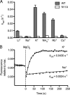
Activation of NFeoBSt by monovalent cations. A, kcat values for wild-type and N11A NFeoBSt in the presence of various monovalent cations. All of the GTP hydrolysis rates were measured in 200 mm salt under Vmax conditions using a colorimetric-based phosphate detection assay. Cations are listed in order of increasing ionic radius: Li+ (76 pm), Na+ (102 pm), K+ (138 pm), NH4+ (144 pm), Rb+ (152 pm), and Cs+ (169 pm). The errors bars are the standard deviations from three independent experiments, with those for N11A too small to be visualized. B, single-turnover hydrolysis of GTP in the presence of K+ and Na+, as measured by intrinsic tryptophan fluorescence from Trp31. 70 μm NFeoBSt was incubated with 30 μm GTP, and then nucleotide binding and hydrolysis was initiated with 1 mm MgCl2 (time 0, dotted line). The drop in fluorescence upon MgCl2 addition is caused by the reorientation of Trp31 in Switch I. As GTP is hydrolyzed, the fluorescence increases as Trp31 adopts its GDP-bound conformation. The kcat values are calculated by fitting the data to a first order exponential association function.

References

    1. Cachero T. G., Morielli A. D., Peralta E. G. (1998) Cell 93, 1077–1085 - PubMed
    1. Finlin B. S., Correll R. N., Pang C., Crump S. M., Satin J., Andres D. A. (2006) J. Biol. Chem. 281, 23557–23566 - PubMed
    1. Schaafsma D., Roscioni S. S., Meurs H., Schmidt M. (2008) Cell Signal. 20, 1705–1714 - PubMed
    1. Karnoub A. E., Weinberg R. A. (2008) Nat. Rev. Mol. Cell Biol. 9, 517–531 - PMC - PubMed
    1. Vetter I. R., Wittinghofer A. (2001) Science 294, 1299–1304 - PubMed

Publication types

MeSH terms

LinkOut - more resources