. 2010 May 7;285(19):14617-27.
doi: 10.1074/jbc.M109.085456. Epub 2010 Mar 10.

AMP-activated protein kinase antagonizes pro-apoptotic extracellular signal-regulated kinase activation by inducing dual-specificity protein phosphatases in response to glucose deprivation in HCT116 carcinoma

Affiliations

AMP-activated protein kinase antagonizes pro-apoptotic extracellular signal-regulated kinase activation by inducing dual-specificity protein phosphatases in response to glucose deprivation in HCT116 carcinoma

Min-Jung Kim et al. J Biol Chem. .

Erratum in

Abstract

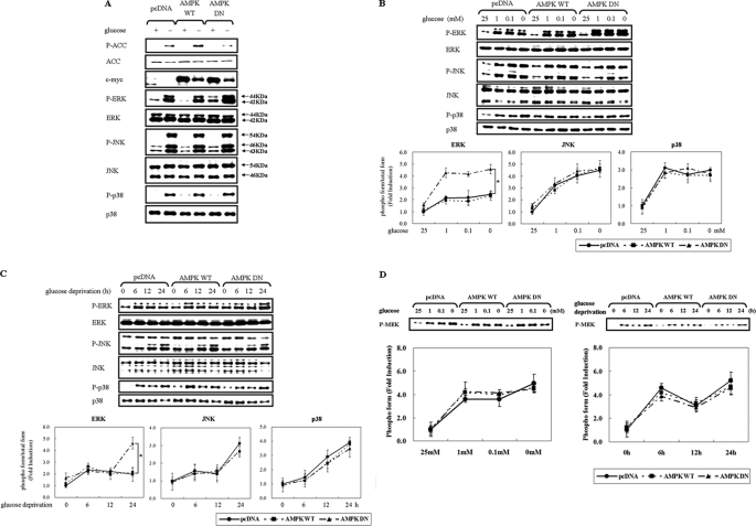
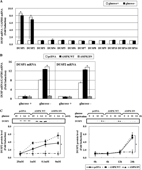
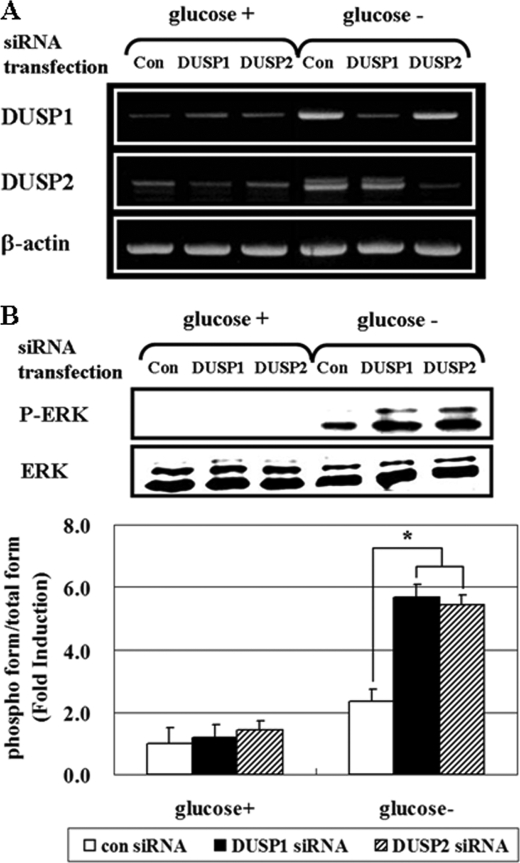
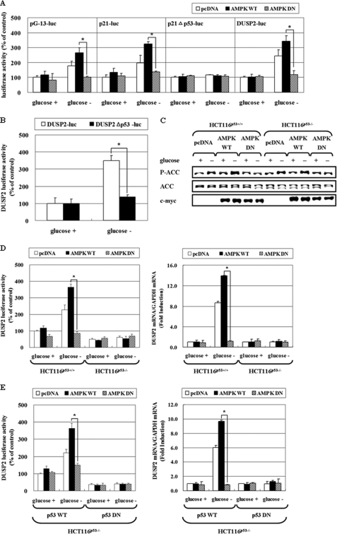
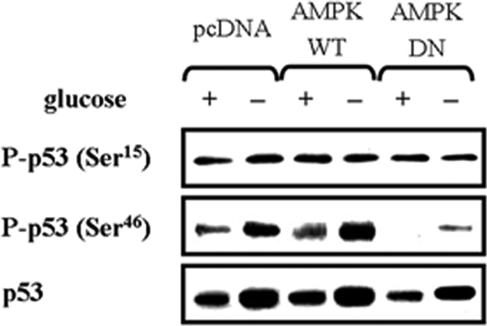
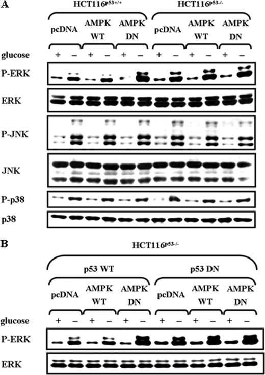
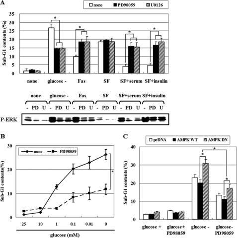
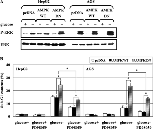
Mitogen-activated protein kinase (MAPK) pathways are involved in the regulation of cellular responses, including cell proliferation, differentiation, cell growth, and apoptosis. Because these responses are tightly related to cellular energy level, AMP-activated protein kinase (AMPK), which plays an essential role in energy homeostasis, has emerged as another key regulator. In the present study, we demonstrate a novel signal network between AMPK and MAPK in HCT116 human colon carcinoma. Glucose deprivation activated AMPK and three MAPK subfamilies, extracellular signal-regulated kinase (ERK), c-Jun NH(2)-terminal kinase (JNK), and p38 MAPK. Under these conditions, inhibition of endogenous AMPK by expressing a dominant-negative form significantly potentiated ERK activation, indicating that glucose deprivation-induced AMPK is specifically antagonizing ERK activity in HCT116 cells. Moreover, we provide novel evidence that AMPK activity is critical for p53-dependent expression of dual-specificity phosphatase (DUSP) 1 & 2, which are negative regulators of ERK. Notably, ERK exhibits pro-apoptotic effects in HCT116 cells under glucose deprivation. Collectively, our data suggest that AMPK protects HCT116 cancer cells from glucose deprivation, in part, via inducing DUSPs, which suppresses pro-apoptotic ERK, further implying that a signal network between AMPK and ERK is a critical regulatory point in coupling the energy status of the cell to the regulation of cell survival.


Figures

FIGURE 1.
FIGURE 1.
Inhibition of AMPK potentiates glucose deprivation-induced ERK activation in a MEK-independent manner. HCT116p53+/+ cells were transfected with pcDNA or c-myc-tagged pAMPK-WT or pAMPK-DN expression vector for 24 h and exposed to glucose deprivation for 24 h (A), the indicated concentration of glucose for 24 h (B, D), or glucose-free medium for the indicated time period (C, D). Under these conditions, the phosphorylated form of the indicated protein (P-ACC, P-ERK, P-JNK, P-p38, P-MEK) and the total form (ACC, ERK, JNK, p38, c-myc) was examined via Western blot analyses using specific antibodies. Experiments were repeated four times with similar results, and a representative result is shown. Each band was analyzed and quantified by densitometer. The data are expressed as the means ± S.E. (*, p < 0.05; compared with the indicated groups).
FIGURE 2.
FIGURE 2.
AMPK activation is critical for DUSP induction in response to glucose deprivation. A, HCT116p53+/+ cells were incubated in glucose-depleted medium for 24 h. Then, the mRNA levels of 12 DUSP members were determined by real-time PCR analysis, and the fold induction is expressed as a ratio of each DUSP: GAPDH mRNA. B, HCT116p53+/+ cells were transfected with pcDNA, pAMPK-WT, and pAMPK-DN expression vectors for 24 h, and maintained under glucose deprivation for 24 h. The amount of DUSP1 and -2 and GAPDH mRNA was evaluated by real-time PCR analysis. Results are the means ± S.E. for six determinations. C, HCT116p53+/+ cells were transfected with pcDNA, pAMPK-WT, and pAMPK-DN expression vectors for 24 h, and then were exposed to medium containing the indicated concentrations of glucose for 24 h (left panel) or incubated in glucose-free medium for the indicated times (right panel). The amount of DUSP2 protein was determined by Western blot analysis, and its level was quantified by densitometer. The data are expressed as the means ± S.E. (*, p < 0.05; compared with the indicated groups.) glucose+, 25 mm glucose; glucose-, 0 mm glucose.
FIGURE 3.
FIGURE 3.
The activation of DUSP2 promoter by glucose deprivation requires AMPK. A, HCT116p53+/+ cells were co-transfected with a luciferase reporter plasmid containing DUSP promoter (pDUSP2-luc) and pcDNA, pAMPK-WT, or pAMPK-DN at a 1:1 ratio. After 24 h, cells were exposed to glucose depleted-medium for 24 h, and then luciferase activity was measured. B, mouse embryonic fibroblasts with AMPKα-deleted (AMPKα−/−) or wild type (AMPKα+/+) were co-transfected with pDUSP2-luc and an expression vector for green fluorescence protein for 24 h, and then exposed to glucose-free medium for 24 h. Then cell lysates were subjected to the luciferase activity assay or the intensity of fluorescence was measured. The data represent means ± S.E. for six determinations. (*, p < 0.05; compared with the indicated groups.)
FIGURE 4.
FIGURE 4.
Down-regulation of DUSP1 and DUSP2 enhances ERK activity. After transfection with siRNA for control, DUSP1, or DUSP2 for 24 h, HCT116p53+/+ cells were maintained in the absence of glucose for 24 h. Then, the mRNA level of DUSP1 and 2 was determined by RT-PCR (A), and phosphoactivated ERK (P-ERK), and its total form (ERK) were determined by Western blot analysis and compared via densitometer (B). Experiments were repeated four times with similar results, and a representative result is shown. (*, p < 0.05; compared with the indicated groups.)
FIGURE 5.
FIGURE 5.
AMPK increases DUSP2 promoter activity via p53 activation. A, HCT116p53+/+ cells were co-trasnfected with pcDNA, pAMPK-WT, or pAMPK-DN expression vectors, and a luciferase reporter containing a promoter with 13 p53 responsive elements (pG-13-luc), a wild-type p21cip1/waf1 promoter (p21-luc), p21 promoter with the p53 binding site deleted (p21cip1/waf1Δp53-luc), or pDUSP2-luc for 24 h. B, HCT116p53+/+ cells were transfected with a luciferase reporter with a wild-type DUSP2 promoter (pDUSP2-luc) or a DUSP2 promoter with the p53 binding site deleted (pDUSP2Δp53-luc) for 24 h. After exposure to glucose-depleted medium for 24 h, cell lysates were subjected to a luciferase activity assay. The data represent means ± S.E. for six determinations. C and D, HCT116p53+/+ and HCT116p53−/− cells were transiently co-transfected with pcDNA, pAMPK-WT, or pAMPK-DN expression vector, and pDUSP2-luc with a 1:1 ratio. After 24 h transfection, cells were exposed to glucose deprivation for 24 h, and then phosphorylation level of ACC and total ACC was measured by Western blot (C). Also, the luciferase activity assay (left panel) or mRNA level of DUSP2 was determined (right panel) (D). E, HCT116p53−/− cells were co-transfected with pDUSP2-luc and/or pcDNA, pAMPK-WT, pAMPK-DN, p53-WT, or p53-DN (p53-R175D) expression vectors at a 1:1:1 ratio. After 24 h transfection, cells were exposed to glucose deprivation for 24 h, and then luciferase activity (left panel) or mRNA level of DUSP2 (right panel) was measured. The data represent means ± S.E. for six determinations. (*, p < 0.05; compared with the indicated groups.)
FIGURE 6.
FIGURE 6.
AMPK regulates DUSP1 mRNA expression via p53 activation in response to glucose deprivation. A, HCT116p53+/+ and HCT116p53−/− cells were transfected with pcDNA, pAMPK-WT, or pAMPK-DN expression vectors for 24 h, and then exposed to glucose-depleted medium for 24 h. Then, the mRNA level of DUSP1 was determined. B, under identical conditions as Fig. 5E (right panel), the mRNA level of DUSP1 was determined. The data represent means ± S.E. for six determinations. (*, p < 0.05; compared with the indicated groups.)
FIGURE 7.
FIGURE 7.
AMPK is required for p53 phosphorylation at Ser46, but not at Ser15, following glucose deprivation. HCT116p53+/+ cells were transfected with pcDNA, pAMPK-WT, or pAMPK-DN expression vectors for 24 h, and then incubated in glucose-depleted medium for 24 h. The phosphorylation level of p53 at Ser15 and Ser46 was analyzed by Western blot. Experiments were repeated three times with similar results, and a representative result is shown.
FIGURE 8.
FIGURE 8.
AMPK negatively regulates ERK phosphorylation in a p53-dependent manner. A, HCT116p53+/+ and HCT116p53−/− cells were transfected with pcDNA, pAMPK-WT, or pAMPK-DN expression vectors for 24 h, and then incubated in glucose depleted-medium for 24 h. B, HCT116p53−/− cells were co-transfected with pcDNA, pAMPK-WT, pAMPK-DN, and p53-WT, p53-DN (p53-R175D) expression vectors at a 1:1 ratio for 24 h, and then incubated in glucose-depleted medium for 24 h. Then, the phosphoactive form and total form of ERK, JNK, and p38 MAPK were analyzed by Western blot. Experiments were repeated three times with similar results, and a representative result is shown.
FIGURE 9.
FIGURE 9.
ERK has contrasting effects depending on the stimulus, and AMPK protects cells from glucose deprivation-induced apoptosis via suppressing pro-apoptotic ERK. A, HCT116p53+/+ cells were pretreated with two different MEK inhibitors, PD98059 (25 μm) or U0126 (10 μm), for 30 min, and then cells were exposed for 24 h to glucose-deprived medium, Fas (CD95/Apo-1)-activating mouse anti-Fas monoclonal antibody (Fas), 10% serum, or insulin (100 nm) following 12 h of serum starvation. Cells were then subjected to fluorescence-activated cell scanning analysis for the percentage of nuclei containing subdiploid amounts of DNA (sub-G1 fraction) (upper panel) and phosphoactivated ERK (P-ERK) was measured (lower panel). B, HCT116p53+/+ cells were exposed to a medium containing the indicated concentrations of glucose for 24 h with or without 25 μm PD98059 and then analyzed for apoptosis via fluorescence-activated cell scanning analysis. C, HCT116p53+/+ cells were transfected with pcDNA, pAMPK-WT, or pAMPK-DN expression vectors for 24 h and maintained under glucose deprivation for 24 h with or without PD98059 (25 μm). The sub-G1 fraction of the cells was measured as a percentage. Experiments were repeated three times with similar results, and a representative result is shown. (*, p < 0.05; compared with the indicated groups.)
FIGURE 10.
FIGURE 10.
Inhibition of AMPK activation by glucose deprivation promotes pro-apoptotic ERK in various human cancer cell lines. A, HepG2 and AGS cells were transfected with pcDNA, pAMPK-WT, or pAMPK-DN expression vectors for 24 h, and then incubated in glucose-depleted medium for 24 h. Then, the phosphoactive form (P-ERK) and total form of ERK (ERK) were analyzed by Western blot. Experiments were repeated three times with similar results and a representative result is shown. B, HepG2 and AGS cells were transfected with pcDNA, pAMPK-WT, or pAMPK-DN expression vectors for 24 h and maintained under glucose deprivation for 24 h with or without PD98059 (25 μm). Percentages of sub-G1 cells via FACS analysis are indicated. Experiments in triplicate were repeated three times. (*, p < 0.05; compared with the indicated groups.)

Similar articles

Cited by

References

    1. Hardie D. G., Carling D., Carlson M. (1998) Annu. Rev. Biochem. 67, 821–855 - PubMed
    1. Shackelford D. B., Shaw R. J. (2009) Nat. Rev. Cancer 9, 563–575 - PMC - PubMed
    1. Inoki K., Ouyang H., Zhu T., Lindvall C., Wang Y., Zhang X., Yang Q., Bennett C., Harada Y., Stankunas K., Wang C. Y., He X., MacDougald O. A., You M., Williams B. O., Guan K. L. (2006) Cell 126, 955–968 - PubMed
    1. Jones R. G., Plas D. R., Kubek S., Buzzai M., Mu J., Xu Y., Birnbaum M. J., Thompson C. B. (2005) Mol. Cell 18, 283–293 - PubMed
    1. Xia Z., Dickens M., Raingeaud J., Davis R. J., Greenberg M. E. (1995) Science 270, 1326–1331 - PubMed

Publication types

MeSH terms