. 2010 May 21;285(21):16275-85.
doi: 10.1074/jbc.M110.102749. Epub 2010 Mar 10.

Trp-26 imparts functional versatility to human alpha-defensin HNP1

Affiliations

Trp-26 imparts functional versatility to human alpha-defensin HNP1

Gang Wei et al. J Biol Chem. .

Abstract

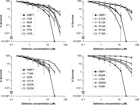
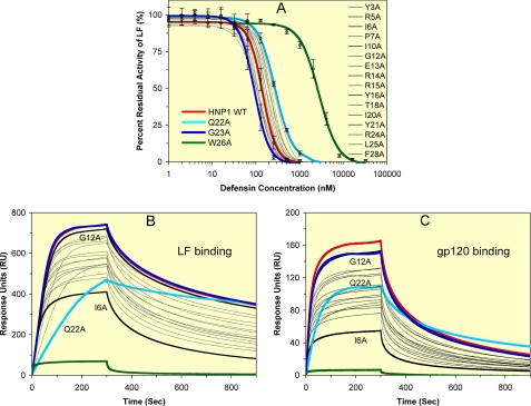
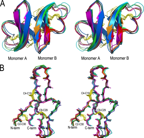
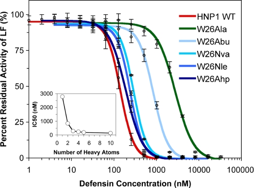
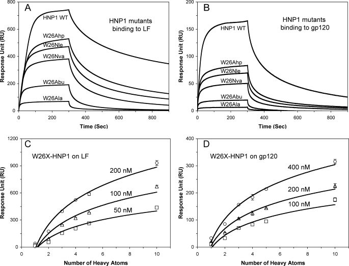
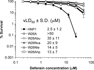
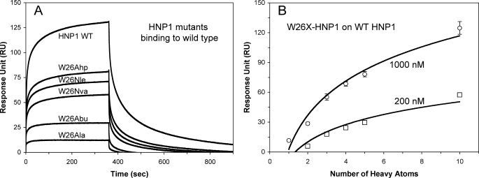
We performed a comprehensive alanine scan of human alpha-defensin HNP1 and tested the ability of the resulting analogs to kill Staphylococcus aureus, inhibit anthrax lethal factor, and bind human immunodeficiency virus-1 gp120. By far, the most deleterious mutation for all of these functions was W26A. The activities lost by W26A-HNP1 were restored progressively by replacing W26 with non-coded, straight-chain aliphatic amino acids of increasing chain length. The hydrophobicity of residue 26 also correlated with the ability of the analogs to bind immobilized wild type HNP1 and to undergo further self-association. Thus, the hydrophobicity of residue 26 is not only a key determinant of the direct interactions of HNP1 with target molecules, but it also governs the ability of this peptide to form dimers and more complex quaternary structures at micromolar concentrations. Although all defensin peptides are cationic, their amphipathicity is at least as important as their positive charge in enabling them to participate in innate host defense.


Figures

FIGURE 1.
FIGURE 1.
Survival curves of S. aureus ATCC 29213 (MicroBioLogics) exposed to HNP1 and its 19 Ala-scan analogs. Strains were exposed to the peptides at concentrations varying from 0.195 to 50 μm. Except for wild type HNP1, each curve is the mean of triplicate experiments, where the error bars represent the S.D. of the measurements. The virtual colony counting assay tests a maximum of six peptides on one 96-well plate, including wild type HNP1 as a control on every plate to ensure internal consistency. The data of wild type HNP1 are presented as averages of 12 independent measurements ±S.D. Because points equivalent to zero survival cannot be plotted on a logarithmic scale, the complete killing achieved by 25 μm wild type HNP1 appears on the x axis, below the scaled portion of the results. A Student's t test was used to calculate the p values for statistical significance of wild type HNP1 versus selective defensin analogs: p = 0.00046 for R5A, p = 0.00076 for R14A, p = 0.0073 for E13A, p = 0.00068 for R15A, p = 0.00016 for R24A, p = 0.00011 for W26A, and p = 0.00063 for F28A.
FIGURE 2.
FIGURE 2.
Inhibition and/or binding of LF and HIV-1 gp120 by wild type HNP1 and its 19 Ala-scan analogs. A, shown is inhibition of LF activity by different concentrations of defensin. The data are the averages of three independent enzyme kinetic measurements, except for HNP1, of which the inhibition curve was obtained from 24 separate assays. For clarity, only the inhibition curves of HNP1, Q22A-HNP1, G23A-HNP1, and W26A-HNP1 are highlighted in color thick lines with error bars. WT, wild type. B and C, binding kinetics of defensins, each at 100 nm, on immobilized LF (2500 RUs) or gp120 (2830 RUs) as determined by SPR. In addition to the four color-coded defensins, HNP1 (red), Q22A-HNP1 (cyan), G23A-HNP1 (blue), and W26A-HNP1 (green), G12A-HNP1 and I6A-HNP1 are highlighted in black.
FIGURE 3.
FIGURE 3.
Structural alignment of Cα traces of dimers and monomers of HNP1 analogs. A, shown is a stereo view of backbone-superimposed dimers of wild type HNP1 (light green), I6A-HNP1 (magenta), Y16A-HNP1 (cyan), Y21A-HNP1 (gray), Q22A-HNP1 (orange), R24A-HNP1 (red), and F28A-HNP1 (blue). B, shown is a stereo view of backbone-superimposed monomers of HNP1 and the six Ala-substituted analogs. Disulfide bonds are represented by yellow sticks.
FIGURE 4.
FIGURE 4.
Native disulfide linkages shown in a NOESY spectrum of W26A-HNP1. The Cys-9—Cys-29 linkage is observed through long-range connectivities with the side chain of Glu-13. The Cys-2—Cys-30 linkage is established through weak cross-strand NOEs between the CδH or CϵH of Phe-28 (7.05 ppm) and the CβH of both Cys-2 and Cys-30.
FIGURE 5.
FIGURE 5.
Inhibition of LF activity by different concentrations of W26X-HNP1 analogs. The data are the averages of three independent enzyme kinetic measurements. For comparison, wild type (WT) HNP1 and W26A-HNP1 are also plotted.
FIGURE 6.
FIGURE 6.
Binding of LF and gp120 by W26X-HNP1 analogs. A and B, shown are binding kinetics of defensins, each at 100 nm, on immobilized LF (2500 RUs) and gp120 (2830 RUs) as determined by SPR. C and D, shown are plots of RU values at 300 s of association of W26X-HNP1 analogs at three different concentrations versus the number of heavy atoms (non-H atoms) in the side chain of residue 26 in HNP1. The data of HNP1 and W26A-HNP1 are included for comparison.
FIGURE 7.
FIGURE 7.
Bactericidal activity of HNP1, W26A-HNP1, and W26X-HNP1 analogs (from 0. 195 to 50 μm) against S. aureus ATCC 29213. Each curve is the mean of three independent experiments, where the error bars represent the S.D. of the measurements. Points scored as 0 survival could not be plotted. vLD50, virtual LD50.
FIGURE 8.
FIGURE 8.
Crystal structures of W26Abu-HNP1 and W26Ahp-HNP1. A, shown is a stereo view of superimposed backbone structures of HNP1 (green), W26Abu-HNP1 (slate), and W26Ahp-HNP1 (orange) dimers. B, shown is a close-up view of the dimer interfaces in monomer A (left panel) and monomer B (right panel). The residues and disulfide bonds at the dimer interface are highlighted as sticks. H-bonds that stabilize the Trp-26 conformation in wild type HNP1 are shown as black dashes.
FIGURE 9.
FIGURE 9.
Self-association of HNP1, W26A-HNP1, and W26X-HNP1 analogs on wild type HNP1 surface. A, shown are binding kinetics of the defensins at 1 μm each on 285 RUs of immobilized HNP1. B, shown are plots of RU values at 300 s of association of the defensins at 200 nm and 1 μm versus the number of heavy atoms (non-H atoms) in the side chain of residue 26 in HNP1. The RU values are the average readings from three separate experiments performed at room temperature. WT, wild type.

Similar articles

Cited by

References

    1. Zasloff M. (2002) Nature 415, 389–395 - PubMed
    1. Habermann E. (1972) Science 177, 314–322 - PubMed
    1. Steiner H., Hultmark D., Engström A., Bennich H., Boman H. G. (1981) Nature 292, 246–248 - PubMed
    1. Zasloff M. (1987) Proc. Natl. Acad. Sci. U.S.A. 84, 5449–5453 - PMC - PubMed
    1. Selsted M. E., Ouellette A. J. (2005) Nat. Immunol. 6, 551–557 - PubMed

Publication types

MeSH terms