. 2010 May 7;285(19):14777-90.
doi: 10.1074/jbc.M109.093708. Epub 2010 Mar 10.

Mutant huntingtin fragments form oligomers in a polyglutamine length-dependent manner in vitro and in vivo

Affiliations

Mutant huntingtin fragments form oligomers in a polyglutamine length-dependent manner in vitro and in vivo

Justin Legleiter et al. J Biol Chem. .

Abstract

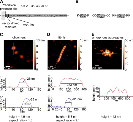
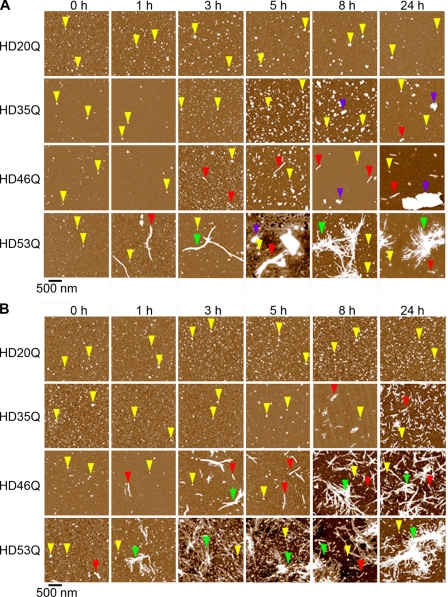
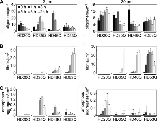
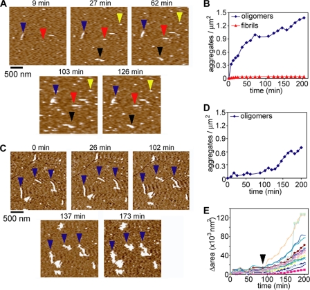
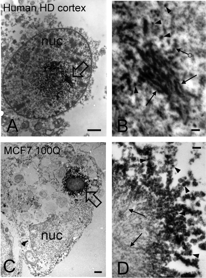
Huntington disease (HD) is caused by an expansion of more than 35-40 polyglutamine (polyQ) repeats in the huntingtin (htt) protein, resulting in accumulation of inclusion bodies containing fibrillar deposits of mutant htt fragments. Intriguingly, polyQ length is directly proportional to the propensity for htt to form fibrils and the severity of HD and is inversely correlated with age of onset. Although the structural basis for htt toxicity is unclear, the formation, abundance, and/or persistence of toxic conformers mediating neuronal dysfunction and degeneration in HD must also depend on polyQ length. Here we used atomic force microscopy to demonstrate mutant htt fragments and synthetic polyQ peptides form oligomers in a polyQ length-dependent manner. By time-lapse atomic force microscopy, oligomers form before fibrils, are transient in nature, and are occasionally direct precursors to fibrils. However, the vast majority of fibrils appear to form by monomer addition coinciding with the disappearance of oligomers. Thus, oligomers must undergo a major structural transition preceding fibril formation. In an immortalized striatal cell line and in brain homogenates from a mouse model of HD, a mutant htt fragment formed oligomers in a polyQ length-dependent manner that were similar in size to those formed in vitro, although these structures accumulated over time in vivo. Finally, using immunoelectron microscopy, we detected oligomeric-like structures in human HD brains. These results demonstrate that oligomer formation by a mutant htt fragment is strongly polyQ length-dependent in vitro and in vivo, consistent with a causative role for these structures, or subsets of these structures, in HD pathogenesis.


Figures

FIGURE 1.
FIGURE 1.
Experimental systems. A, a schematic representation is shown of the GST-htt exon 1 fusion proteins used in this study (HD20Q, HD35Q, HD46Q, or HD53Q). These fusion proteins contain a PreScission protease site located between GST and the htt fragment (not drawn to scale). Cleavage of the GST initiates the aggregation reaction. B, schematics of the synthetic polyQ peptides used in this study are shown. Flanking lysine residues were added to increase solubility. Representative AFM images and aggregate profiles that compare basic dimensions of oligomers (C), fibrils (D), and amorphous aggregates of HD53Q (E) are shown. Height profiles under each image are indicated by colored lines. By using a combination of height and aspect ratio, relative populations of aggregates types can be distinguished and quantified in a heterogeneous mixture.
FIGURE 2.
FIGURE 2.
Oligomers and fibrils formed by a mutant htt fragment depend on polyQ length and concentration. AFM images demonstrate the aggregation of mutant htt fragments with polyQ repeats of 20Q, 35Q, 46Q, or 53Q. Incubations with protein concentrations of 2 μm (A) and 30 μm (B) were imaged at different time points after the addition of protease. Examples of oligomers, fibrils, fibril clusters, and large amorphous aggregates are indicated by yellow, red, green, and blue arrows, respectively. Although oligomers were observed for HD20Q, fibrillar aggregates were not. For mutant htt fragments with longer polyQ repeats, heterogeneous mixtures of aggregate types appeared at later times. The appearance of fibrillar aggregates of HD35Q was concentration-dependent. At 2 μm, HD35Q began to form large amorphous aggregates but at 30 μm HD35Q formed a large number of short fibrils. At late times, fibrils and fibril bundles were the dominant morphology for HD46Q and HD53Q, with fibrils appearing earlier for HD53Q at both concentrations.
FIGURE 3.
FIGURE 3.
Aggregate species of mutant htt fragments change temporally in a polyQ length- and concentration-dependent manner. Quantification of aggregate types observed in AFM images for incubations HD20Q, HD35Q, HD46Q, and HD53Q at 2 μm and 30 μm. The numbers of oligomers (A), fibrils (B), and amorphous (C) aggregates per μm2 were calculated for all images in Fig. 2. The appearance of fibrils occurred earlier for longer polyQ repeat lengths. Although HD20Q formed oligomers at both concentrations, fibrils were not observed. For HD35Q, a peak population of oligomers preceded the formation of amorphous aggregates (2 μm) or fibrils (30 μm). A peak population of oligomers also preceded the appearance of fibrils of HD46Q at both concentrations. As fibrils of HD53Q appeared early in the experiment, a peak oligomer population preceding the appearance of fibrils was only observed if the kinetics of aggregation reactions were decreased (see supplemental Fig. 2).
FIGURE 4.
FIGURE 4.
Oligomers formed by mutant htt fragments are highly heterogeneous in size. Based on corrected volume measurements (see supplemental Fig. 3) and the molecular mass of each htt exon 1 fragment, the numbers of molecules per oligomer observed for 2 μm incubations of HD20Q, HD35Q, HD46Q, or HD53Q were calculated for each time point of aggregation from Fig. 2. The plots are color-coded such that darker colors represent a greater abundance of oligomers composed of that number of molecules. Black arrows indicate where ∼400 kDa oligomers would be observed, and the maximum size in kDa is represented in the plots as marked. Based on this analysis, htt exon 1 fragments form a mixture of oligomers ∼400 kDa and larger, similar to what was observed by SDS-AGE in vitro and in vivo (see Fig. 7).
FIGURE 5.
FIGURE 5.
A mutant htt fragment forms oligomers in solution. A, monomeric preparations of HD53Q were imaged in solution on mica at a concentration of 2 μm. The yellow arrows indicate oligomers that are stable throughout the experiment. The red arrows indicate an oligomer that coalesced into a fibril. The blue and black arrows indicate fibrils that appeared without a clear oligomeric precursor. B, the number of oligomers and fibrils per unit area was determined as a function of time. Oligomers appeared on the surface by 9 min with a steady increase in abundance over time. The vast majority of these oligomers did not further aggregate into fibrillar structures. Very short (<200 nm in length) fibrillar structures also appeared within 9 min. Although the number of fibrils did increase slightly, they were far less abundant than oligomers. C, HD53Q was aggregated to a fibrillar state, deposited on mica, and imaged in solution. Next, a monomeric preparation of HD53Q (2 μm) was injected into the fluid cell. D, the number of oligomers per unit area was determined as a function of time, illustrating that fewer oligomers formed in the presence of fibrils. E, the surface area occupied by individual fibrils was monitored as a function of time. Initially, fibrils appeared to elongate predominately at the fibril ends, resulting in a slight increase in occupied surface area. However, ∼88 min (marked by black arrowhead) after the addition of HD53Q monomer, branching appeared on several fibrils, increasing the rate of change in occupied surface area. The fibrils then appeared to accumulate soluble HD53Q as the fibrils expanded in all directions (also see supplemental Movie 1).
FIGURE 6.
FIGURE 6.
The synthetic polyQ peptide KK-Q32-KK forms oligomers with faster kinetics than a mutant htt fragment. A, in situ AFM experiments were performed with 5 μm HD53Q and KK-Q32-KK. B, the number of oligomeric and fibrillar aggregates formed by HD53Q and KK-Q32-KK was measured as a function of time. For KK-Q32-KK, the number of oligomers peaked (∼80 min) before dissipating, as the number of fibrils continued to increase. Large amorphous aggregates of KK-Q32-KK also appeared. Under these conditions, HD53Q only formed oligomers, which steadily increased in number as a function of time. C, ex situ AFM image of KK-Q32-KK sampled 5 h after incubation at 5 μm demonstrating that the peptide formed a mixture of aggregates consisting of oligomers (yellow arrows) and two distinct types of fibrils: narrower fibrils (∼1–2 nm tall, red arrows) and taller fibrils (∼5–7 nm tall, blue arrows).
FIGURE 7.
FIGURE 7.
A mutant htt fragment forms oligomers in a polyQ length-dependent manner in neurons and brains. A, SDS-AGE with Western analysis indicates that a mutant htt fragment (HD53Q) forms oligomers that migrate as a smear above ∼400 kDa when detected with an anti-htt antibody (EM48). B and C, a mutant htt fragment forms oligomers in a polyQ length-dependent manner in ST14A cells similar in apparent size to those formed under cell-free conditions. Note that oligomers formed by HD97Q-GFP are less heterogeneous in size than those formed with shorter polyQ lengths but that the intensity of oligomers as detected by antibody reactivity increases as a function of time in ST14A cells. D, a filter-trap retardation assay shows that the total amount of SDS-insoluble aggregates increases as a function of polyQ length and time in ST14A cells that express a mutant htt fragment. E and F, a mutant htt fragment forms oligomers in total brain homogenates from R6/2 mice that are similar in apparent size to those formed under cell-free conditions. Note that oligomers formed by a mutant htt fragment with 220Q are less heterogeneous in size than those formed with shorter polyQ lengths and that the intensity of antibody reactivity also is significantly decreased in R6/2 mice with 220Q. WT, wild type. G and H, SDS-AGE with Western analysis on total brain homogenates show that a mutant htt fragment forms oligomers that significantly increase as a function of time in R6/2 mice with 110Q. I, SDS-AGE with Western analysis on the supernatant fraction of a total brain homogenate after centrifugation shows that a mutant htt fragment forms soluble oligomers that increase as a function of time in R6/2 mice with 110Q. *, p < 0.05; ***, p < 0.0001 (one-way analysis of variance).
FIGURE 8.
FIGURE 8.
Mutant htt forms oligomers in HD brains. Mutant htt was detected with EM using the immunoperoxidase method and a primary antibody to htt1–17. Analysis was performed with a transmission electron microscope (JEOL 100CX). The peroxidase reaction product appears as electron dense label. A and B, electron micrographs are from a cortical neuron in human postmortem HD brain. The CAG repeats were 17 and 69. Note that the labeled inclusion occupies a large area at the center of the nucleus (nuc). The composition of the inclusion is highly heterogeneous consisting of granular/oligomeric and fibrillar structures. Stacks of labeled fibrils are present at the upper part of the inclusion and along the right at the open arrow, which is magnified in B. In B, note the presence of small, labeled granular/oligomeric structures of different sizes (arrowheads) that are similar in lateral dimensions to those observed by AFM analysis of mutant htt fragments in vitro. Fibrils (arrows) and structures that resemble beads on a string (ringed arrow) are also indicated. C and D, electron micrographs of an MCF7 cell transfected with Flag-htt1–969-100Q. In C, a large cytoplasmic inclusion appears adjacent to the nucleus (nuc) at the open arrow. A portion of the inclusion is shown at higher magnification in D. The inclusion consists of a core of unlabeled fibrils (arrows) and a periphery of immunoreactive granular/oligomeric structures (arrowheads). Some of the granules/oligomers form linear arrays (“beads on a string”) similar to that observed in the human HD brain (B, ringed arrow). Scale bars on left panels = 1 μm and on right panels = 100 nm.
FIGURE 9.
FIGURE 9.
A model for the misfolding and aggregation of a mutant htt fragment. A native monomer can continuously misfold to sample distinct monomeric conformers. The relative number and stability of these conformers may be polyQ length-dependent. Some of these misfolded monomers potentially can lead to off-pathway aggregates, such as large annular aggregates. Others can form globular oligomeric aggregates. The size and stability of oligomeric aggregates can vary widely and are also polyQ length-dependent. These oligomers can further self-assemble or accumulate into other aggregate morphologies, such as small annular aggregates composed of oligomeric subunits, large amorphous aggregates, and fibrils. In this model a major structural transition must occur within an oligomer to initiate fibril elongation. Once this transition occurs, oligomers and monomers can directly accumulate on the fibril, causing fibril elongation. Many of these higher order aggregates can accumulate together to form large aggregates that make up inclusions.

References

    1. The Huntington's Disease Collaborative Research Group (1993) Cell 72, 971–983 - PubMed
    1. DiFiglia M., Sapp E., Chase K. O., Davies S. W., Bates G. P., Vonsattel J. P., Aronin N. (1997) Science 277, 1990–1993 - PubMed
    1. Zoghbi H. Y., Orr H. T. (2000) Annu Rev. Neurosci. 23, 217–247 - PubMed
    1. Penney J. B., Jr., Vonsattel J. P., MacDonald M. E., Gusella J. F., Myers R. H. (1997) Ann. Neurol. 41, 689–692 - PubMed
    1. Snell R. G., MacMillan J. C., Cheadle J. P., Fenton I., Lazarou L. P., Davies P., MacDonald M. E., Gusella J. F., Harper P. S., Shaw D. J. (1993) Nat. Genet. 4, 393–397 - PubMed

Publication types

MeSH terms

LinkOut - more resources