. 2010 May 7;285(19):14475-85.
doi: 10.1074/jbc.M109.084400. Epub 2010 Mar 10.

The cancer/testis antigen CAGE with oncogenic potential stimulates cell proliferation by up-regulating cyclins D1 and E in an AP-1- and E2F-dependent manner

Affiliations

The cancer/testis antigen CAGE with oncogenic potential stimulates cell proliferation by up-regulating cyclins D1 and E in an AP-1- and E2F-dependent manner

Elaine Por et al. J Biol Chem. .

Abstract

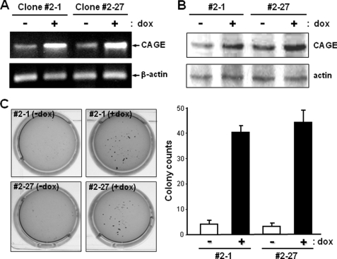
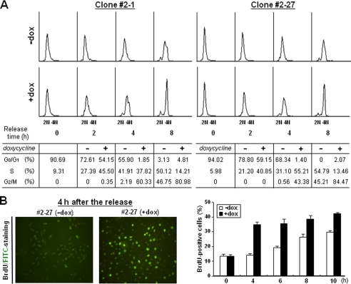
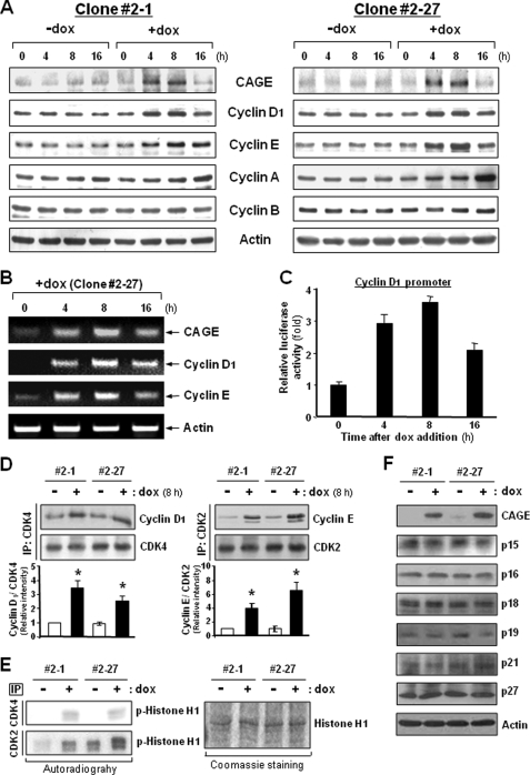
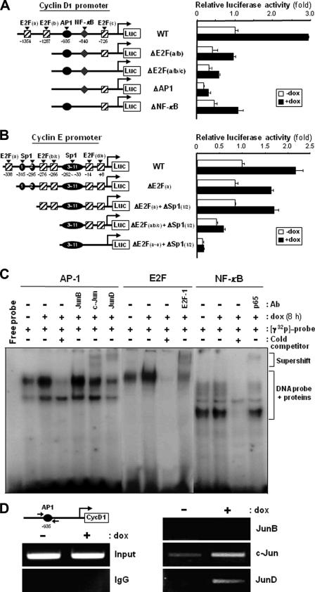
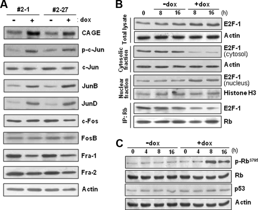
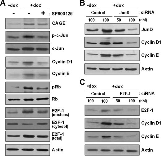
A cancer/testis antigen, CAGE, is widely expressed in various cancer tissues and cancer cell lines but not in normal tissues except the testis. In the present study, ectopic expression of CAGE in fibroblast cells resulted in foci formation, suggesting its cell-transforming ability. Using stable HeLa transfectant clones with the tetracycline-inducible CAGE gene, we found that CAGE overexpression stimulated both anchorage-dependent and -independent cell growth in vitro and promoted tumor growth in a xenograft mouse model. Cell cycle analysis showed that CAGE augments the levels of cyclin D1 and E, thereby activating cyclin-associated cyclin-dependent kinases and subsequently accelerating the G(1) to S progression. Moreover, increased cyclin D1 and E levels in CAGE-overexpressing cells were observed even in a growth arrested state, indicating a direct effect of CAGE on G(1) cyclin expression. CAGE-induced expression of cyclins D1 and E was found to be mediated by AP-1 and E2F-1 transcription factors, and among the AP-1 members, c-Jun and JunD appeared to participate in CAGE-mediated up-regulation of cyclin D1. CAGE overexpression also enhanced retinoblastoma phosphorylation and subsequent E2F-1 nuclear translocation. In contrast, small interfering RNA-mediated knockdown of CAGE suppressed the expression of G(1) cyclins, activation of AP-1 and E2F-1, and cell proliferation in both HeLa cervical cancer cells and Malme-3M melanoma cells. These results suggest that the cancer/testis antigen CAGE possesses oncogenic potential and promotes cell cycle progression by inducing AP-1- and E2F-dependent expression of cyclins D1 and E.


Figures

FIGURE 1.
FIGURE 1.
Generation of stable tetracycline-inducible CAGE transfectant clones of HeLa cells and their colony-forming ability in soft agar. A and B, Tet-On sublines of HeLa cells were transfected with CAGE cDNA subcloned into a pTRE2 tetracycline-inducible expression vector. The resultant stable transfectant clones were analyzed for CAGE expression in the absence or presence of doxycycline (dox) (1 μg/ml) by RT-PCR (A) and immunoblotting (B) analyses. C, stable tetracycline-inducible CAGE transfectant clones 2-1 and 2-27 suspended in 0.3% soft agar were cultured in the absence or presence of doxycycline for 21 days. After crystal violet staining, colonies with a diameter larger than 1 mm were counted directly. Values are the means ± S.D. of triplicate cultures.
FIGURE 2.
FIGURE 2.
In vivo and in vitro growth rate of HeLa cells with tetracycline-inducible CAGE. A and B, stable tetracycline-inducible CAGE transfectant clones (106 cells) were inoculated subcutaneously into athymic nude mouse. Following inoculation 10 mice were divided into two groups, with one group devoid of treatment and the other group administered doxycycline (dox) (1 μg/ml). At the onset of tumor formation, tumor diameter was measured every other day with calipers (A). At 35 days after inoculation, the mice were euthanized, and tumor weight was measured (B). Values are the means ± S.D. of tumors from five mice. CAGE abundance in representative primary tumors (P) from each mouse was assessed by immunoblotting with an antibody to CAGE (inset in B). C, tetracycline-inducible CAGE transfectant clones were seeded at a low density and cultured in the absence or presence of doxycycline (1 μg/ml), and cell numbers were counted directly every 24 h over the course of 6 days. For delayed overexpression of CAGE, doxycycline was added to the culture medium of untreated cells 3 days after initial seeding of the cells (arrows above Δ+dox). *, p < 0.01 versusdox at day 6 in culture.
FIGURE 3.
FIGURE 3.
Cell cycle progression in CAGE-overexpressing HeLa cells. A, tetracycline-inducible CAGE transfectant clones were synchronized by double thymidine for G1/S block. After treatment with or without doxycycline for 2 h, synchronized cells were released in complete medium supplemented with or without doxycycline for the indicated time periods. After cells were labeled with propidium iodide, DNA content was analyzed by flow cytometry. Shown are representative histograms and the means of cell cycle phase distribution from three independent experiments. B, cells synchronized by double thymidine block were released in the absence or presence of doxycycline (dox) for the indicated time periods. After BrdUrd treatment, cells were stained with fluorescein isothiocyanate (FITC)-conjugated anti-BrdUrd antibody and viewed under a fluorescence microscope. Shown are representative images and the means ± S.D. of BrdUrd-positive cell counts in triplicate cultures.
FIGURE 4.
FIGURE 4.
Expression levels of cyclins and kinase activities of CDKs in CAGE-overexpressing HeLa cells. A and B, asynchronized HeLa cells transfected with the tetracycline-inducible CAGE were cultured in the absence or presence of doxycycline (dox) for the indicated time periods. Expression levels of cyclins D1, E, A, and B were examined by immunoblotting (A) and RT-PCR (B) analyses. C, following transfection with a control pGL3 reporter vector or vector containing cyclin D1 promoter DNA, CAGE transfectant cells were treated with doxycycline for the indicated time periods. Promoter activity was assessed by a luciferase assay. D, the amount of cyclins D1 and E associated with CDK4 and CDK2, respectively, were assessed by immunoprecipitation (IP) followed by immunoblotting analysis. *, p < 0.05 versusdox. E, immunoprecipitates with anti-CDK4 and anti-CDK2 antibodies were subjected to an in vitro kinase assay using histone H1 as a substrate. F, CDK inhibitor proteins were examined for their cellular levels by immunoblotting analysis.
FIGURE 5.
FIGURE 5.
Functional involvement of AP-1 and E2F binding sites within cyclin D1 and E gene promoters in CAGE-induced G1 cyclin expression. A and B, human wild-type (WT) promoters of the cyclin D1 (A) and E (B) genes and the mutant promoters for E2F, AP-1, NF-κB, and Sp1 binding sites were fused to a luciferase reporter gene of the pGL3 vector. Following transient transfection with pGL3 vector containing wild-type or mutant promoters of cyclin D1 and E genes, the tetracycline-inducible CAGE transfectant clone 2-27 was treated with or without doxycycline (dox) for 8 h, and a dual luciferase assay was performed. Normalized luciferase activity ± S.D. of triplicate experiments are plotted. C, double-stranded oligonucleotides containing AP-1, E2F, or NF-κB recognition sequences were 32P-labeled and incubated with nuclear extracts of CAGE transfectant cells treated with or without doxycycline. Specific DNA binding activities of AP-1, E2F, and NF-κB factors were determined by an electrophoretic mobility shift assay in the absence or presence of an 8-fold excess of cold competitors. Anti-JunB, anti-c-Jun, anti-JunD, anti-E2F-1 and anti-NF-κB antibodies were used for supershift analysis. D, CAGE transfectant cells treated with or without doxycycline were fixed with formaldehyde and then harvested and sonicated to shear DNA. Following incubation with anti-JunB, anti-c-Jun, or anti-JunD antibodies or normal mouse IgG, the immunocomplex was subjected to 25 PCR cycles using primers specific to the AP-1 binding region of the human cyclin D1 promoter.
FIGURE 6.
FIGURE 6.
Levels of AP-1 proteins, Rb phosphorylation, and nuclear E2F in CAGE-overexpressing cells. A, protein levels of Jun and Fos subfamily members of AP-1 factors were examined in tetracycline-inducible CAGE transfectant HeLa cells by immunoblotting analysis after doxycycline (dox) treatment for 8 h. The phosphorylation level of c-Jun was evaluated by using an antibody specific to the phospho-Ser-63/Ser-73 residues of c-Jun. B, CAGE transfectant clonal cells (#2-27) were treated with or without doxycycline for the indicated time periods, and E2F-1 protein levels in the total cell lysate, the cytosolic and nuclear fractions, and the Rb immunoprecipitate (IP) were examined for by immunoblotting analysis. C, the phosphorylation level of Rb in cells (#2-27) treated with or without doxycycline was assessed by immunoblotting analysis using an antibody specific to Rb phosphorylated at the Ser-795 residue.
FIGURE 7.
FIGURE 7.
Ablation of CAGE-induced G1 cyclin expression by an inhibitor of JNK and siRNAs for JunD and E2F-1. A, the tetracycline-inducible CAGE transfectant clone (#2-27) of HeLa cells was pretreated with a JNK inhibitor, SP600125, for 4 h and further incubated in the absence or presence of doxycycline (dox) for 8 h. The levels of cyclin D1 and E proteins, phospho-c-Jun, phospho-Rb, and cytosolic and nuclear E2F-1 proteins were assessed by immunoblotting analysis. B and C, the clonal cells (#2-27) were transfected with control and JunD (B) or E2F-1 (C) siRNAs. At 48 hours after transfection, cells were treated with or without doxycycline for 8 h and analyzed for protein levels of cyclins D1 and E.
FIGURE 8.
FIGURE 8.
CAGE siRNA-mediated suppression of G1 cyclin expression, Jun AP-1 protein expression/activation, E2F-1 nuclear translocation, and cell proliferation in human cancer cell lines. A and B, protein levels of CAGE, cyclins D1 and E, and JunD and the phosphorylation level of c-Jun (A) and Rb (B) were determined in CAGE siRNA-transfected HeLa and Malme-3M cells. Cont, control. C, the level of cytosolic and nuclear E2F-1 was assessed by immunoblotting analysis after subcellular fractionation. D and E, cell proliferation was measured by MTS assay upon siRNA knockdown of CAGE in HeLa and Malme-3M cells as described under “Experimental Procedures.” *, p < 0.01 versus −doxycycline at day 4 in culture.

Similar articles

Cited by

References

    1. Boon T., Old L. J. (1997) Curr. Opin. Immunol. 9, 681–683 - PubMed
    1. Scanlan M. J., Gure A. O., Jungbluth A. A., Old L. J., Chen Y. T. (2002) Immunol. Rev. 188, 22–32 - PubMed
    1. Scanlan M. J., Simpson A. J., Old L. J. (2004) Cancer Immun. 4, 1. - PubMed
    1. Zendman A. J., Ruiter D. J., Van Muijen G. N. (2003) J. Cell. Physiol. 194, 272–288 - PubMed
    1. van der Bruggen P., Traversari C., Chomez P., Lurquin C., De Plaen E., Van den Eynde B., Knuth A., Boon T. (1991) Science 254, 1643–1647 - PubMed

Publication types

MeSH terms