

Fetal weight normograms for singleton pregnancies in a Jordanian population
- PMID: 20220263
- PMCID: PMC2855064
- DOI: 10.4103/0256-4947.60519
Fetal weight normograms for singleton pregnancies in a Jordanian population
Abstract
Background and objectives: Estimated intrauterine fetal weight (EIUFW) is important for reducing prenatal mortality and morbidity through early detection of faltering growth. Our objectives were to develop patterns of ultrasonically determined EIUFW by gestational age, for normal singleton pregnancies, and to assess the effect of a number of variables on EIUFW.
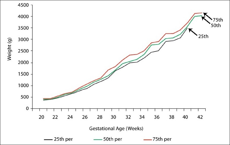
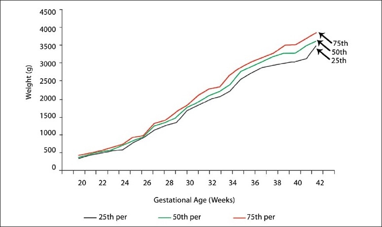
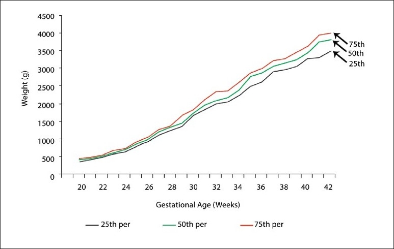
Methods: Ultrasonically, EIUFW was obtained from 600 pregnant women who were at 20 to 42 weeks of gestation (WG). EIUFW was categorized into low weight and normal weight using the tenth and twentieth percentile as the cut-off points. Logistic regression was used to calculate the odds ratio and their 95% confidence limits for a number of risk factors hypothesized to be associated with low fetal weight. EIUFW percentiles (twenty-fifth, fiftieth, and seventy-fifth), by gestational age and sex, were calculated for singleton pregnancies.
Results: Up to 32 WG there was no statistically significant difference between male and female fetuses in EIUFW. Between 32 and 39 WG males had significantly (P< .05) higher fetal weight than females. Charts of ultrasonically determined EIUFW by gestational age and sex for singleton pregnancies were created. A number of variables were significantly associated with EIUFW, such as pregnancy weight gain, maternal hemoglobin level, frequency of antenatal visits, smoking status, and fetal sex.
Conclusion: Weight gain during pregnancy should be encouraged for pregnant mothers who gain less than one kilogram per month in the second and third trimester. A prospective study on a national representative sample in Jordan is needed to generate our own standards for fetal growth.
Figures
Similar articles
-
Unisex vs sex-specific estimated fetal weight charts for fetal growth monitoring: a population-based study.Am J Obstet Gynecol MFM. 2022 Jan;4(1):100527. doi: 10.1016/j.ajogmf.2021.100527. Epub 2021 Nov 8. Am J Obstet Gynecol MFM. 2022. PMID: 34763120
-
Fetal growth restriction and small for gestational age as predictors of neonatal morbidity: which growth nomogram to use?Am J Obstet Gynecol. 2023 Dec;229(6):678.e1-678.e16. doi: 10.1016/j.ajog.2023.06.035. Epub 2023 Jun 20. Am J Obstet Gynecol. 2023. PMID: 37348779
-
Prediction of large-for-gestational-age neonate by routine third-trimester ultrasound.Ultrasound Obstet Gynecol. 2019 Sep;54(3):326-333. doi: 10.1002/uog.20377. Epub 2019 Jul 23. Ultrasound Obstet Gynecol. 2019. PMID: 31236963
-
The World Health Organization fetal growth charts: concept, findings, interpretation, and application.Am J Obstet Gynecol. 2018 Feb;218(2S):S619-S629. doi: 10.1016/j.ajog.2017.12.010. Am J Obstet Gynecol. 2018. PMID: 29422204 Review.
-
Should twin-specific growth charts be used to assess fetal growth in twin pregnancies?Am J Obstet Gynecol. 2022 Jul;227(1):10-28. doi: 10.1016/j.ajog.2022.01.027. Epub 2022 Jan 31. Am J Obstet Gynecol. 2022. PMID: 35114185 Review.
References
-
- McCormick MC, Brooks-Gunn J, Workman-Daniels K, Turner J, Peckham GJ. The health and developmental status of very low-birth-weight children at school age. JAMA. 1992;267:2204–8. - PubMed
-
- Bennett BB. Shoulder dystocia: An obstetric emergency. Obstet Gynecol Clin North Am. 1999;26:445–58. - PubMed
-
- Phillips DI. Birth weight and the future development of diabetes: A review of the evidence. Diabetes Care. 1998;21:150–5. - PubMed
MeSH terms
LinkOut - more resources
Full Text Sources