. 2010 Mar 11;464(7286):237-242.
doi: 10.1038/nature08852.

Somatic sex identity is cell autonomous in the chicken

Affiliations

Somatic sex identity is cell autonomous in the chicken

D Zhao et al. Nature. .

Abstract

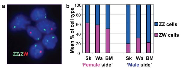
In the mammalian model of sex determination, embryos are considered to be sexually indifferent until the transient action of a sex-determining gene initiates gonadal differentiation. Although this model is thought to apply to all vertebrates, this has yet to be established. Here we have examined three lateral gynandromorph chickens (a rare, naturally occurring phenomenon in which one side of the animal appears male and the other female) to investigate the sex-determining mechanism in birds. These studies demonstrated that gynandromorph birds are genuine male:female chimaeras, and indicated that male and female avian somatic cells may have an inherent sex identity. To test this hypothesis, we transplanted presumptive mesoderm between embryos of reciprocal sexes to generate embryos containing male:female chimaeric gonads. In contrast to the outcome for mammalian mixed-sex chimaeras, in chicken mixed-sex chimaeras the donor cells were excluded from the functional structures of the host gonad. In an example where female tissue was transplanted into a male host, donor cells contributing to the developing testis retained a female identity and expressed a marker of female function. Our study demonstrates that avian somatic cells possess an inherent sex identity and that, in birds, sexual differentiation is substantively cell autonomous.


Figures

Figure 1
Figure 1
Image of gynandromorph bird (G1) ISA Brown bird where the right side has female characteristics and left side has male characteristics (white colour and larger wattle, breast musculature & spur).
Figure 2
Figure 2
Male and female cells in gynandromorph birds. a. FISH analysis of sex chromosomes in gynandromorph blood cells. Interphase nuclei prepared from cultured blood cells from gynandromorph G1 hybridised according to standard FISH procedures with probes specific to both the W and Z chromosome (XhoI repeat on W chromosome, and Z chromosome BAC containing the VLDL receptor gene identified by screening the HGMP chicken BAC library). Erythrocytes hybridised with probes for Z-chromosome (GREEN) and W-chromosome (RED). Cells contain either two Z-chromosomes or one Z and one W-chromosome. b. Mean relative proportions of ZZ and ZW cells in tissues from male and female sides of gynandromorph birds. Average percentage of ZW cells and ZZ cells (Supplementary Table 2) in three tissues from phenotypically female side and from phenotypically male side of three gynandromorph birds. Tissues from the sides that appear female contain more ZW (female) than ZZ (male) cells, while tissues from the sides that appear male are composed predominantly of ZZ cells. Sk=skin, Wa=wattle, BM=breast muscle.
Figure 3
Figure 3
Sexually dimorphic expression in early chick embryos. a. Expression of Faf in male and female embryos prior to development of genital ridge/gonads. Whole-mount ISH showing expression of Faf (purple colour) in embryos at 18hrs, 48hrs and 72hrs of development (H&H stages 4, 14 & 20). Faf is clearly expressed throughout the female embryos at all developmental stages and not expressed in male embryos. Faf is not expressed in extra-embryonic tissues of the female. The Faf transcript is encoded by the genomic DNA complementary to the intergenic regions between copies of the W-chromosome repeat gene Wpkci / HINTW , and transcribed in the opposite orientation. m: male, f:female. b. Expression of novel chicken miRNA (gga-mir-2954). Expression in whole embryos at 48hrs (H&H 14) and 72 hrs (H&H 20) of development. This miRNA is clearly expressed in a sexually dimorphic fashion at stages prior to the sexual differentiation of the gonads. This miRNA matches sequence present in chicken Z-chromosome BAC clones AC192757 and AC187119. Loading Control= U6 RNA.
Figure 4
Figure 4
Expression of male and female markers in chimeric gonads. a. Generation of chimeras. I) Schematic illustrating transplantation of presumptive mesoderm from GFP-expressing embryo to non-GFP embryo at day 2. II) Image of mesonephros and gonads from chimeric embryo at day 9 showing donor contribution to left gonad (g), mesonephros (m) and mullerian duct (md). b. Expression of female and male markers in embryonic gonads. Expression of aromatase (AROM) in ovary and anti-Mullerian hormone (AMH) in testis at day 9 is shown by IHC. c. Integration of GFP-donor cells into host gonads. Panels in first column show low magnification view of sections through host gonads and illustrate the extent of donor cell contribution. Remaining panels show higher magnification views of highlighted areas. Using IHC, donor cells are marked by GFP (green) while expression of AMH and aromatase are shown in red. Fourth column shows merged image of images from second and third columns. In same sex chimeras, GFP-expressing donor cells co-localise with AMH-expressing and aromatase-expressing cells in host testis and ovary respectively ( yellow/orange in fourth column). In mixed-sex chimeras GFP-expressing donor cells do not co-localise with AMH or aromatase. m: mesonephros, o: ovary, and t: testis. d. Retention of female donor phenotype in mixed-sex chimeras. IHC showing expression of AMH (red in top row of panels) and aromatase (red in bottom row of panels) in neighbouring sections from the gonad of a female:male (donor:host) chimera. Donor contribution is illustrated by GFP (green) expression. Third column shows merged image of images in first and second columns. Regions containing a significant host contribution (defined by bottom bracket in top row of panels) formed sex-cord like structures and expressed AMH. Female donor cells were not incorporated into AMH expressing sex cords, as shown by lack of GFP and AMH co-localisation. Regions composed primarily of female donor cells (defined by top bracket) behaved as ovarian-like tissue and expressed aromatase, as shown by co-localisation of GFP and aromatase (yellow/orange). Scale bars represent 100 μm. IHC was performed following standard procedures. Primary antibodies were (1:100) goat anti-human AMH (Santa Cruz Biotechnology), (1:200) mouse anti-human cytochrome P450 aromatase (AbD Serotec) and (1:250) rabbit anti-GFP conjugated to Alexa Fluor 488 (Invitrogen). Secondary antibodies were conjugated to Alexa Fluor 594 (Invitrogen).
Figure 5
Figure 5
A novel mechanism of sex determination in the chicken. A sexual identity is genetically imposed on the male and female chicken soma at fertilisation and is the major factor in determining the adult sexual phenotype. At the appropriate stage in development, the sexually-dimorphic transcripts underlying the male / female identity trigger expression in the genital ridge of the gene cascade responsible for testis / ovary development. The gonads have limited effects on the sexual phenotype. In contrast, in mammals, gonadal fate is dependent on transient expression of the testis-determining SRY gene in the indifferent early gonad. The mammalian gonads have a major influence on the sexual phenotype.

Comment in

References

    1. Lillie FR. Sex-determination and sex-differentiation in mammals. Proc. Natl. Acad. Sci. USA. 1917;3:464–470. - PMC - PubMed
    1. Jost A. Hormonal factors in the sex differentiation of the mammalian foetus. Phil. Trans. R. Soc. Lond. B. 1970;259:119–130. - PubMed
    1. Agate RJ, et al. Neural, not gonadal, origin of brain sex differences in a gynandromorph finch. Proc. Natl. Acad. Sci. USA. 2003;100:4873–4878. - PMC - PubMed
    1. Wade J, Arnold AP. Functional testicular tissue does not masculinize development of the zebra finch song system. Proc. Natl. Acad. Sci. USA. 1996;93:5264–5268. - PMC - PubMed
    1. Arnold AP. Sexual differentiation of the zebra finch song system: positive evidence, negative evidence, null hypothesis, and a paradigm shift. J. Neurobiol. 1997;33:572–584. - PubMed

Methods References

    1. Stern CD. Immunocytochemistry of embryonic material. In: Stern CD, Holland PWH, editors. Essential Developmental Biology: A Practical Approach. IRL Press; Oxford, UK: 1993. pp. 193–212.
    1. McQueen HA, et al. CpG islands of chicken are concentrated on microchromosomes. Nature Genetics. 1996;12:321–324. - PubMed
    1. Clinton M, Haines L, Belloir B, Mcbride D. Sexing chick embryos: a rapid and simple protocol. Br. Poultry Sci. 2001;42:134–138. - PubMed
    1. Henrique D, et al. Expression of a Delta homologue in prospective neurons in the chick. Nature. 1995;375:787–790. - PubMed
    1. Clinton M, Miele G, Nandi S, McBride D. Identification of disease markers by differential display: prion disease. In: Liang P, Meade JD, Pardee AB, editors. Differential Display Methods and Protocols. 2nd edition Humana Press; Totowa, NJ: 2006. pp. 157–178. - PubMed

Publication types

Substances