. 2010 Feb;14(1):1-9.
doi: 10.4196/kjpp.2010.14.1.1. Epub 2010 Feb 28.

Mechanical Stretch-Induced Protection against Myocardial Ischemia-Reperfusion Injury Involves AMP-Activated Protein Kinase

Affiliations

Mechanical Stretch-Induced Protection against Myocardial Ischemia-Reperfusion Injury Involves AMP-Activated Protein Kinase

Jia Hao et al. Korean J Physiol Pharmacol. 2010 Feb.

Abstract

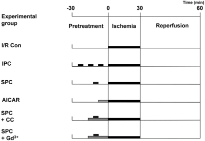
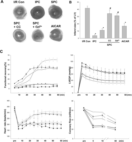
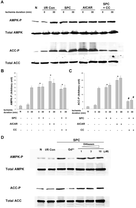
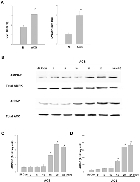
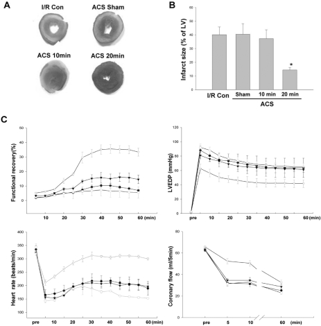
AMP-activated protein kinase (AMPK) protects various tissues and cells from ischemic insults and is activated by many stimuli including mechanical stretch. Therefore, this study investigated if the activation of AMPK is involved in stretch-induced cardioprotection (SIC). Intraventricular balloon and aorto-caval shunt (ACS) were used to stretch rat hearts ex vivo and in vivo, respectively. Stretch preconditioning reduced myocardial infarct induced by ischemia-reperfusion (I/R) and improved post-ischemic functional recovery. Phosphorylation of AMPK and its downstream substrate, acetyl-CoA carboxylase (ACC) were increased by mechanical stretch and ACC phosphorylation was completely blocked by the AMPK inhibitor, Compound C. AMPK activator (AICAR) mimicked SIC. Gadolinium, a blocker of stretch-activated ion channels (SACs), inhibited the stretch-induced phosphorylation of AMPK and ACC, whereas diltiazem, a specific L-type calcium channel blocker, did not affect AMPK activation. Furthermore, SIC was abrogated by Compound C and gadolinium. The in vivo stretch induced by ACS increased AMPK activation and reduced myocardial infarct. These findings indicate that stretch preconditioning can induce the cardioprotection against I/R injury, and activation of AMPK plays an important role in SIC, which might be mediated by SACs.

Keywords: AMP-activated protein kinase; Cardioprotection; Ischemia-reperfusion; Stretch.


Figures

Fig. 1
Fig. 1
Protocols for each experimental group, showing the components and the time course of various treatments. All the hearts underwent 30 minutes sustained ischemia followed by 1 hour reperfusion. Prior to the sustained ischemia, the hearts were assigned to each experimental group with specific treatment; no treatment, three cycles of a 5 minutes ischemia, 5 minutes stretch, 10 minutes AICAR treatment, CC with 5 minutes stretch, and Gd3+ with 5 minutes stretch. I/R Con, ischemia-reperfusion control; SPC, stretch preconditioning; IPC, ischemia preconditioning; AICAR, 5-Aminoimidazole-4-carboxamide-1-β-D-ribofuranoside; CC, compound C; Gd3+, gadolinium.
Fig. 2
Fig. 2
Effects of stretch preconditioning in ex vivo hearts. (A) After each experimental protocol, slices of heart were stained with TTC and (B) normalized infarct size was calculated and compared. There was no significant difference among IPC, SPC and AICAR group. All data are means±SEM of N≥6 in each experimental group. *p<0.05 compared with I/R Con group, #p<0.05 compared with SPC group. I/R Con, ischemia-reperfusion control; SPC, stretch preconditioning; IPC, ischemia preconditioning; AICAR, 5-Aminoimidazole-4-carboxamide-1-β-D-ribofuranoside; CC, compound C; Gd3+, gadolinium. (C) Functional recovery of ex vivo stretched hearts. During post-ischemic period, cardiac function was evaluated in terms of 4 parameters; cardiac functional recovery, Left ventricular end-diastolic pressure (LVEDP), heart rate, and coronary blood flow. The changes of 4 parameters were traced during 60 minutes post-ischemia-reperfusion period. There was no significant difference among IPC, SPC and AICAR group. All data are means±SEM of N≥6 in each experimental group. ●, ischemia-reperfusion control; ○, ischemic preconditioning; ▾, stretch preconditioning; ▿, AICAR; ■, stretch+Compound C; □, stretch+gadolinium.
Fig. 3
Fig. 3
The phosphorylation of AMPK and ACC in the ex vivo hearts All the hearts underwent 30 minutes sustained ischemia. (A) The phosphorylation of AMPK and ACC was analyzed with immunoblot and (B, C) semi-quantitative densitometry. The phosphorylation level before and after the ischemia with various pretreatment was compared. (D) Effects of gadolinium and diltiazem on AMPK and ACC phosphorylation induced by SPC. Hearts were treated with gadolinium (10 µM) or diltiazem (1, 3, and 10 µM) during SPC. Results are representative from 6 independent experiments and means±SEM. *p<0.05 compared with I/R Con group. #p<0.05 compared with SPC group. N, normal control; I/R Con, ischemia-reperfusion control; SPC, stretch preconditioning; CC, compound C; Gd3+, gadolinium.
Fig. 4
Fig. 4
Phosphorylation of AMPK and ACC after aorto-caval shunt. (A) Left ventricular wall stress caused by aorto-caval shunt (ACS). Central venous pressure (CVP) and left ventricular end-diastolic pressure (LVEDP) were monitored after ACS. *p<0.05, N=6. (B) After ACS was fitted to the rats, phosphorylation of AMPK and ACC was analyzed at various time points with immunoblot and (C, D) semi-quantitative densitometry. Results are representative from 6 independent experiments. *p<0.05 compared with I/R Con group. I/R C, ischemia-reperfusion control; ACS, aorto-caval shunt.
Fig. 5
Fig. 5
Effects of stretch preconditioning on the infarct size and the functional recovery of in vivo rat hearts. (A) Infarct size was measured using TTC staining after 30 minutes ischemia and 1 hour reperfusion and (B) normalized infarct size was calculated and compared. All data are means±SEM of N≥6 in each experimental group. *p<0.05 compared with I/R Con group. I/R Con, ischemia-reperfusion control; ACS Sham, sham-operated hearts; ACS, aorto-caval shunt. (C) Functional recovery of in vivo stretched rat hearts. Cardiac function was evaluated in terms of 4 parameters; cardiac functional recovery, Left ventricular end-diastolic pressure (LVEDP), heart rate, and coronary blood flow. The changes of 4 parameters were traced during 60 minutes post-ischemic reperfusion period. All data are means±SEM of N≥6 in each experimental group. *p<0.05 compared with control group. ●, ischemia-reperfusion control; ○, Sham; ▾, 10 minutes ACS; ▿, 20 minutes ACS; ACS, aorto-caval shunt.

Similar articles

Cited by

References

    1. Das DK, Maulik N. Cardiac genomic response following preconditioning stimulus. Cardiovasc Res. 2006;70:254–263. - PubMed
    1. Shintani-Ishida K, Nakajima M, Uemura K, Yoshida K. Ischemic preconditioning protects cardiomyocytes against ischemic injury by inducing GRP78. Biochem Biophys Res Commun. 2006;345:1600–1605. - PubMed
    1. Kim YH, Kim CH, Kim GT, Kim IK, Park JW, Kim MS. Involvement of adenosine in cardioprotective effect of catecholamine preconditioning in ischemia-reperfused heart of rat. Korean J Physiol Pharmacol. 1998;2:753–761.
    1. Jiao JD, Garg V, Yang B, Hu K. Novel functional role of heat shock protein 90 in ATP-sensitive K+ channel-mediated hypoxic preconditioning. Cardiovasc Res. 2008;77:126–133. - PubMed
    1. Ovize M, Kloner RA, Przyklenk K. Stretch preconditions canine myocardium. Am J Physiol. 1994;266:H137–H146. - PubMed