. 2010 Mar 8;5(3):e9561.
doi: 10.1371/journal.pone.0009561.

Damage of guinea pig heart and arteries by a trioleate-enriched diet and of cultured cardiomyocytes by oleic acid

Affiliations

Damage of guinea pig heart and arteries by a trioleate-enriched diet and of cultured cardiomyocytes by oleic acid

Josef Krieglstein et al. PLoS One. .

Abstract

Background: Mono-unsaturated fatty acids (MUFAs) like oleic acid have been shown to cause apoptosis of cultured endothelial cells by activating protein phosphatase type 2C alpha and beta (PP2C). The question arises whether damage of endothelial or other cells could be observed in intact animals fed with a trioleate-enriched diet.

Methodology/principal findings: Dunkin-Hartley guinea pigs were fed with a trioleate-enriched diet for 5 months. Advanced atherosclerotic changes of the aorta and the coronary arteries could not be seen but the arteries appeared in a pre-atherosclerotic stage of vascular remodelling. However, the weight and size of the hearts were lower than in controls and the number of apoptotic myocytes increased in the hearts of trioleate-fed animals. To confirm the idea that oleic acid may have caused this apoptosis by activation of PP2C, cultured cardiomyocytes from guinea pigs and mice were treated with various lipids. It was demonstrable that oleic acid dose-dependently caused apoptosis of cardiomyocytes from both species, yet, similar to previous experiments with cultured neurons and endothelial cells, stearic acid, elaidic acid and oleic acid methylester did not. The apoptotic effect caused by oleic acid was diminished when PP2C alpha and beta were downregulated by siRNA showing that PP2C was causally involved in apoptosis caused by oleic acid.

Conclusions/significance: The glycerol trioleate diet given to guinea pigs for 5 months did not cause marked atherosclerosis but clearly damaged the hearts by activating PP2C alpha and beta. The diet used with 24% (wt/wt) glycerol trioleate is not comparable to human diets. The detrimental role of MUFAs for guinea pig heart tissue in vivo is shown for the first time. Whether it is true for humans remains to be shown.


Conflict of interest statement

Competing Interests: The authors have declared that no competing interests exist.

Figures

Figure 1
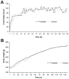
Figure 1. Food consumption and development of the weight of the guinea pigs.
(A) Food uptake measured in calories and (B) body weight in gram of the guinea pigs from both groups.
Figure 2
Figure 2. Levels of the serum lipids after feeding a trioleate-enriched diet for 5 months.
Total cholesterol, LDL cholesterol and HDL cholesterol, but not triglycerides, were significantly increased in the serum of the trioleate-fed guinea pigs. Serum lipids (mg/dl) are given as means ± SD. ***p<0.001, compared with control group.
Figure 3
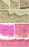
Figure 3. Trioleate-induced changes of the aorta.
Oil red O staining: The aortas from the control diet guinea pigs had no fat inclusions (A). In contrast, the aortas from the trioleate-fed guinea pigs showed some lipid depositions in the subendothelium (B, D) and in the adventitia (C). Furthermore, focal areas with elastin degradation and disorganisation were detected (B, C). HE-staining: The aortas from the control group exhibited a compact structure (E). The media consisted of homogenous layers of thick elastic lamellae and smooth muscle cells. The adventitia was composed of compact connective tissue with interspersed adventitial cells. In comparison, the aortas from the trioleate group exhibited deterioration of the smooth muscle cells and of structural integrity in the media (F. G). The connective tissue of the adventitia was spongy and fewer cells were observed (F). Particularly, in the subendothelial area clusters of foamy smooth muscle cells were found (G). A, C  =  original magnification ×20; B, E, F  =  original magnification ×40.
Figure 4
Figure 4. Size and weight of the guinea pig hearts.
The trioleate-fed guinea pigs show a significantly decreased length (A) and weight (B) of the heart. The ratio of heart weight to body weight (g/kg) was significantly decreased in the trioleate-fed guinea pigs (C). Values are given as means ± SD. ***p<0.001, compared with control group.
Figure 5
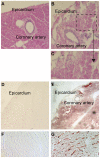
Figure 5. Trioleate-induced damage of guinea pig hearts.
The HE-staining visualized a compact structure of the myocardium of the control-diet group (A). In contrast, trioleate-diet resulted in perivascular fibrosis and a structural deterioration of the myocyte compartment (B, C). Numerous myocytes appeared foamy (B, C). In the coronary arteries of both groups intimal thickening was not observed (A, B, E). Oil red O staining: Lipid accumulations were not found in the control hearts (D, F). In comparison, lipids accumulated throughout the entire myocardium of the trioleate-diet group (E, G). Although massive lipid deposits were detected in the myocardial areas adjacent to coronary arteries, mural lipid deposits were not observed (E). Moreover, lipids accumulated in the interstitial space (G). A, B  =  original magnification ×10; D, E  =  original magnification ×2,5; F, G  =  original magnification ×20.
Figure 6
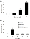
Figure 6. Oleic acid but not stearic acid, elaidic acid or oleic acid methylester damage cardiomyocytes.
Guinea pig cardiomyocytes were incubated with different concentrations of oleic acid and oleic acid induced apoptosis in a dose-dependent manner (A). In contrast, stearic acid, elaidic acid or oleic acid methylester in the concentration range of 200 µM did not stimulate apoptosis (B). Control cardiomyocytes were treated with vehicle only (0.1% DMSO). Apoptotic cells are expressed as percentage of the total number of cells. Means ± SD of 6 experiments are given. ***p<0.001, compared with vehicle-treated control.
Figure 7
Figure 7. Uptake of fatty acids into guinea pig cardiomyocytes.
Guinea pig cardiomyocytes were incubated for 24 h with 200 µM stearic acid (A, D) or 200 µM oleic acid (B, E) and were identified with α-actinin (A, B). Additionally, Nile blue staining showed that both fatty acids were taken up by the cells but, notably, oleic acid-treated cells were partially damaged and stearic acid-treated were not. –  = 10 µm.
Figure 8
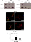
Figure 8. PP2C is causally involved in oleic acid-induced apoptosis in neonatal cardiomyocytes from mice.
Sixteen hours after the onset of transfection, non-transfected as well as transfected cardiomyocytes were treated with 200 µM oleic acid for 4 h. Control cells were incubated with medium plus oligofectamine. PP2C α and β were analyzed by western blot at the beginning and at the end of the experiment (A, B). At both time points, the enzymes in the transfected cells were reduced compared to non-transfected cells. α-Actinin staining (red) demonstrated the typical structure of neonatal cardiomyocytes (C–F). Downregulation of PP2C α and β (green) was observed in these cells (D, F). The cardiomyocytes with reduced PP2C levels were less damaged by oleic acid (G). Apoptotic cells are expressed as percentage of the total number of cells ± SD. ***p<0.001, compared with vehicle-treated control, ## p<0.01, compared with non-transfected cells treated with oleic acid. −  = 10 µm.

Similar articles

Cited by

References

    1. Klumpp S, Selke D, Hermesmeier J. Protein phosphatase type 2C active at physiological Mg2+: stimulation by unsaturated fatty acids. FEBS Lett. 1998;437:229–232. - PubMed
    1. Klumpp S, Selke D, Ahlemeyer B, Schaper C, Krieglstein J. Relationship between protein phosphatase type 2C activity and induction of apoptosis in cultured neuronal cells. Neurochem Int. 2002;41:251–259. - PubMed
    1. Hufnagel B, Dworak M, Soufi M, Mester Z, Zhu Y, et al. Unsaturated fatty acids isolated from human lipoproteins activate protein phospatase type 2Cβ and induce apoptosis in endothelial cells. Atherosclerosis. 2005;180:245–254. - PubMed
    1. Schwarz S, Hufnagel B, Dworak M, Klumpp S, Krieglstein J. Protein phosphatase type 2Cα and 2Cβ are involved in fatty-acid-induced apoptosis of neuronal and endothelial cells. Apoptosis. 2006;11:1111–1119. - PubMed
    1. Klumpp S, Thissen MC, Krieglstein J. Protein phosphatases types 2Cα and 2Cβ in apoptosis. Biochem Soc Trans. 2006;34:1370–1375. - PubMed

Publication types