. 2010 Mar 5;5(3):e9478.
doi: 10.1371/journal.pone.0009478.

Adaptive autoimmunity and Foxp3-based immunoregulation in zebrafish

Affiliations

Adaptive autoimmunity and Foxp3-based immunoregulation in zebrafish

Francisco J Quintana et al. PLoS One. .

Abstract

Background: Jawed vertebrates generate their immune-receptor repertoire by a recombinatorial mechanism that has the potential to produce harmful autoreactive lymphocytes. In mammals, peripheral tolerance to self-antigens is enforced by Foxp3(+) regulatory T cells. Recombinatorial mechanisms also operate in teleosts, but active immunoregulation is thought to be a late incorporation to the vertebrate lineage.

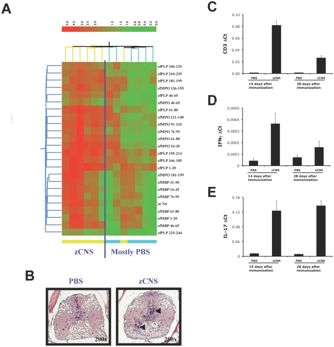
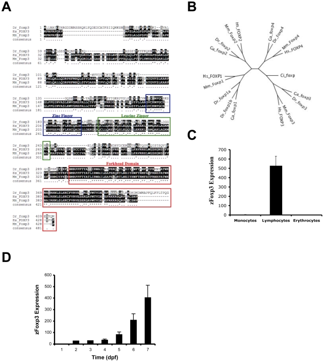
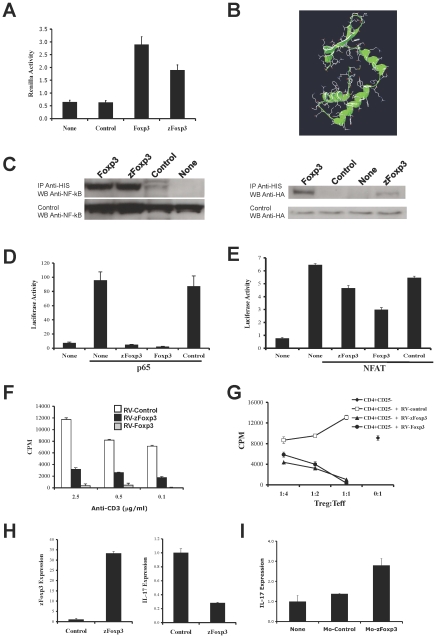
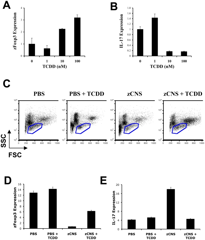
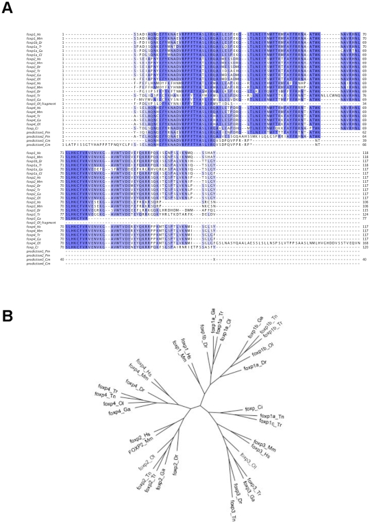
Methods/principal findings: Here we report the characterization of adaptive autoimmunity and Foxp3-based immunoregulation in the zebrafish. We found that zebrafish immunization with an homogenate of zebrafish central nervous system (zCNS) triggered CNS inflammation and specific antibodies. We cloned the zebrafish ortholog for mammalian Foxp3 (zFoxp3) which induced a regulatory phenotype on mouse T cells and controlled IL-17 production in zebrafish embryos.

Conclusions/significance: Our findings demonstrate the acquisition of active mechanisms of self-tolerance early in vertebrate evolution, suggesting that active regulatory mechanisms accompany the development of the molecular potential for adaptive autoimmunity. Moreover, they identify the zebrafish as a tool to study the molecular pathways controlling adaptive immunity.


Conflict of interest statement

Competing Interests: The authors have declared that no competing interests exist.

Figures

Figure 1
Figure 1. Adaptive autoimmunity in zebrafish.
(A) Heatmap depicting the autoantibody response to myelin antigens on day 28 after immunization with zCNS or PBS in CFA. Each column represents a serum sample, color-coded at the bottom to indicate whether it corresponds to a zCNS or a control immunized sample. Only significantly up-regulated antibody reactivities are shown (n = 8, t-test FDR <0.05), according to the colorimetric scale on the right. (B–E) Zebrafish were immunized with zCNS or PBS in CFA and 14 or 28 days later the expression of CD3, IL-17 and IFNγ in brain was measured by real time PCR (mean + s.d. of triplicates) (B–D) or analyzed histologically for the presence of cell infiltrates (E). Two independent experiments produced similar results.
Figure 2
Figure 2. Zebrafish Foxp3 (zFoxp3).
(A) Sequence comparison of putative FoxP3 genes of zebrafish, human and mouse. The stars indicate identity, dashes were introduced for optimal alignment. The zinc finger, leucine zipper and forkhead domains are highlighted with a blue, green or red box, respectively. (B) Radial gene tree showing the Foxp1, Foxp2, Foxp3 and Foxp4 proteins in mammals and fish, where the Ciona intestinalis Foxp sequence is the outgroup. The branch lengths are proportional to the distance between the sequences. Mm, Mus musculus; Hs, Homo sapiens; Dr, Danio rerio; Ga, Gasterosteus aculeatus (stickleback); Ci, Ciona intestinalis. The accession numbers for the amino acid sequences used in the gene tree analysis are as follows: Danio rerio Foxp1a Q08BX8 BC124513; Foxp1b Q2LE08 NM_001039637; Foxp2 Q4JNX5 NM_001030082; Foxp3 annotated (EST CK028390); Foxp4 annotated. Homo sapiens: Foxp1 Q9H334 NM_001012505, Foxp2 O15409 NM_148899, Foxp3 Q9BZS1 NM_014009, Foxp4 Q8IVH2 NM_138457; Mus musculus: Foxp1 P58462 NM_053202, Foxp2 P58463 NM_053242, Foxp3 Q99JB6 NM_054039, Foxp4 Q9DBY0 NM_028767; Ciona intestinalis Foxp Q4H3H6. The amino acid sequence of the apparent stickleback orthologues of Foxp1, Foxp2, Foxp3 and Foxp4 were obtained from Ensembl. (C) Monocytes, lymphocytes and erythrocytes were sorted by FACS and the expression of zFoxP3 was determined by real time PCR (mean + s.d. of triplicates). (D) zFoxp3 and GAPDH were quantified by qPCR on cDNA prepared from zebrafish embryos at different times after fertilization. Two independent experiments produced similar results.
Figure 3
Figure 3. zFoxp3 is a functional homologue of mammalian Foxp3.
(A) Constructs coding for His-labeled zFoxp3 and Renilla-labeled Foxp3 were co-transfected into 293T cells. 24 h later the cells were lysed, zFoxp3 was pulled-down with Ni-Agarose and the renilla luciferase activity in the pellet was quantified. The results are normalized for the total amount of luciferase before precipitation (mean + s.d. of triplicates). Three independent experiments produced similar results. (B) Structure of the forkhead domain of zFoxp3 obtained by homology modeling, based on the structure of the crystallized forkhead domain of Foxp1. (C) 293T cells were co-transfected with His-tagged zFoxp3, Foxp3 and NF-kB or HA-flagged NFAT and 24 hr later the cells were lyzed and immunoprecipitated with antibodies to His antibodies. The precipitates were resolved by PAGE-SDS and detected by western blot with antibodies to NF-kB or HA antibodies. Three independent experiments produced similar results. (D, E) 293T cells were co-transfected with reporter constructs coding for luciferase under the control of a NF-kB or NFAT responsive promoters, and p65 NF-kB or NFAT in the presence of vectors coding for zFoxp3, Foxp3 or control (empty vector). Luciferase activity was normalized to the renilla activity of a co-transfected control (mean + s.d. of triplicates). Four independent experiments produced similar results. (F) MACS-purified CD4+CD25 T-cells were transduced with a bicistronic retrovirus coding for GFP and zFoxp3, Foxp3 or an empty control retrovirus, and the GFP+ population was analyzed for its proliferation upon activation with plate bound antibodies to CD3 (mean cpm or pg/ml + s.d. in triplicate wells) and (G) its suppressive activity on the proliferation and IL-2 and IFNγ secretion of mouse CD4+CD25 T-cells activated with plate-bound antibodies to CD3 (mean cpm or pg/ml + s.d. in triplicate wells). Two independent experiments produced similar results. (H) Fertilized zebrafish eggs were microinjected with a plasmid coding for zFoxp3 or an empty plasmid, and zFoxp3 and IL-17 expression were measured from 6 days old embryos by real time PCR. Two independent experiments produced similar results. (I) Fertilized zebrafish eggs were microinjected with a morpholino oligonucleotides designed to interfere with the translation of zFoxP3 (Mo-zFoxp3) or a 5 bases mismatch control oligonucleotide and IL-17 expression was measured in 5 days old embryos by real time PCR. Two independent experiments produced similar results.
Figure 4
Figure 4. AHR controls zFoxp3 expression.
(A,B) TCDD was added to the water of three-day post-fertilization zebrafish embryos, and 72 h later zFoxp3 (A) and IL-17 (B) expression were determined by real time PCR (mean + s.d. of triplicates normalized to GAPDH expression). Two independent experiments produced similar results. (C) Fourteen days after immunization, kidney cells from PBS or zCNS immunized zebrafish, or TCDD-treated zCNS immunized zebrafish were analyzed by FACS and cells in the lymphocyte fraction (blue gate) were sorted. (D–E) Expression of zFoxp3 and IL-17 measured by real-time PCR in FACS-sorted lymphocytes. Two independent experiments produced similar results.
Figure 5
Figure 5. Evolutionary conservation of the forkhead domains of the FOX family.
(A) Multiple sequence alignment for the forkhead domains of the FOX family. The alignment includes the sequence for Foxp1–4 for Homo sapiens, Mus musculus (Genbank/EMBL sequences) and several of the sequences for the teleosts Danio rerio (Dr), Oryzias latipes (Ol), Takifugu rubripes (Tr) and Gasterosteus aculeatus (Ga) (Ensembl annotation). The partial sequences prediction1_Pm and prediction2_Pm were obtained by assembling sequenced sea lamprey reads. Similarly the sequences prediction3_Cm ad prediction4_Cm are from sequenced reads from the elephant shark genome project (Callorhinchus milii). Although these predicted sequences show characteristic FOXP motifs, as for example the N-terminus motif RPPFTYA, there is strong indication that they are not part of FOXP3 forkhead domains, as for example the motif LIAQAI that in FOXP3 forkhead is replaced by LIRWAI. (B) Radial gene tree for the forkhead domains of the FOX family showing the Foxp1, Foxp2, Foxp3 and Foxp4 proteins in mammals and fish, where the Ciona intestinalis Foxp sequence is the outgroup. The branch lengths are proportional to the distance between the sequences.

References

    1. Pancer Z, Cooper MD. The evolution of adaptive immunity. Annu Rev Immunol. 2006;24:497–518. - PubMed
    1. Honjo T, Nagaoka H, Shinkura R, Muramatsu M. AID to overcome the limitations of genomic information. Nat Immunol. 2005;6:655–661. - PubMed
    1. Bouaziz JD, Yanaba K, Tedder TF. Regulatory B cells as inhibitors of immune responses and inflammation. Immunol Rev. 2008;224:201–214. - PubMed
    1. Hogquist KA, Baldwin TA, Jameson SC. Central tolerance: learning self-control in the thymus. Nat Rev Immunol. 2005;5:772–782. - PubMed
    1. Roncarolo MG, Battaglia M. Regulatory T-cell immunotherapy for tolerance to self antigens and alloantigens in humans. Nat Rev Immunol. 2007;7:585–598. - PubMed

Publication types