. 2010 Mar 5;6(3):e1000799.
doi: 10.1371/journal.ppat.1000799.

Kaposi's sarcoma-associated herpesvirus ORF57 protein binds and protects a nuclear noncoding RNA from cellular RNA decay pathways

Affiliations

Kaposi's sarcoma-associated herpesvirus ORF57 protein binds and protects a nuclear noncoding RNA from cellular RNA decay pathways

Brooke B Sahin et al. PLoS Pathog. .

Abstract

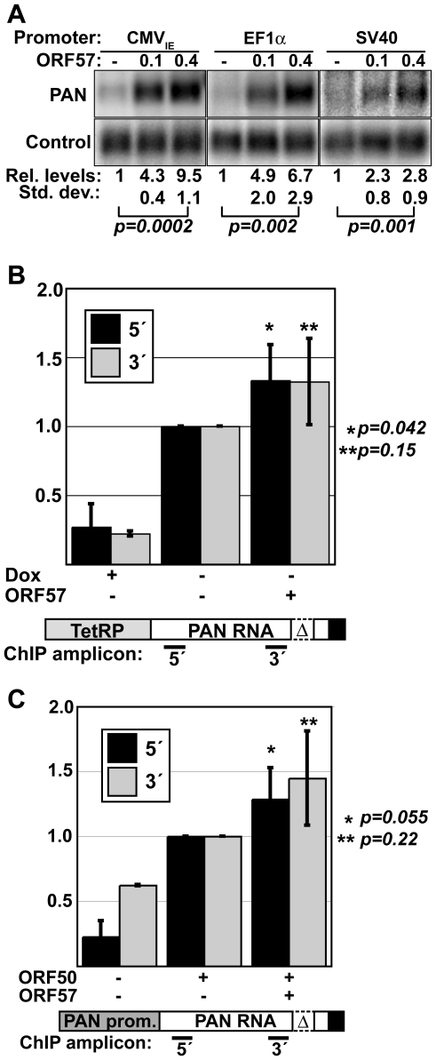
The control of RNA stability is a key determinant in cellular gene expression. The stability of any transcript is modulated through the activity of cis- or trans-acting regulatory factors as well as cellular quality control systems that ensure the integrity of a transcript. As a result, invading viral pathogens must be able to subvert cellular RNA decay pathways capable of destroying viral transcripts. Here we report that the Kaposi's sarcoma-associated herpesvirus (KSHV) ORF57 protein binds to a unique KSHV polyadenylated nuclear RNA, called PAN RNA, and protects it from degradation by cellular factors. ORF57 increases PAN RNA levels and its effects are greatest on unstable alleles of PAN RNA. Kinetic analysis of transcription pulse assays shows that ORF57 protects PAN RNA from a rapid cellular RNA decay process, but ORF57 has little effect on transcription or PAN RNA localization based on chromatin immunoprecipitation and in situ hybridization experiments, respectively. Using a UV cross-linking technique, we further demonstrate that ORF57 binds PAN RNA directly in living cells and we show that binding correlates with function. In addition, we define an ORF57-responsive element (ORE) that is necessary for ORF57 binding to PAN RNA and sufficient to confer ORF57-response to a heterologous intronless beta-globin mRNA, but not its spliced counterparts. We conclude that ORF57 binds to viral transcripts in the nucleus and protects them from a cellular RNA decay pathway. We propose that KSHV ORF57 protein functions to enhance the nuclear stability of intronless viral transcripts by protecting them from a cellular RNA quality control pathway.


Conflict of interest statement

The authors have declared that no competing interests exist.

Figures

Figure 1
Figure 1. ORF57 preferentially enhances the levels of an unstable nuclear RNA.
(A) Top, schematic diagram of the constructs used. Both are driven by the PAN promoter (gray) and have the PAN 3′-end formation signals (black). PAN-Δ79 has the 79-nt ENE sequence deleted. Bottom, representative northern blot showing a dose-dependent response of PAN-WT and PAN-Δ79 to ORF57. Cells were co-transfected with PAN and ORF57 expression plasmids as indicated. Because these constructs use the ORF50-dependent PAN promoter, an ORF50 expression plasmid was also co-transfected. Control panels show signal from a co-transfected plasmid that controls for transfection and loading efficiencies. (B) Quantitation of dose-dependent experiments shown in (A). Values are normalized to the no ORF57 control lanes; error bars show standard deviation (n = 4).
Figure 2
Figure 2. ORF57 protects transcripts from rapid RNA decay.
(A) Top, schematic diagram of TRP-Δ79 construct, which contains the Tet-responsive promoter driving PAN-Δ79. Bottom, representative northern blots showing typical transcription pulse data. The upper panels show data from a two-hour pulse in the presence or absence of ORF57 as indicated. The “-2” lanes are samples taken prior to the pulse. The lower panels show the results from an 18-hr transcription pulse. The asterisks mark the mobility of hyperadenylated transcripts. Cellular 7SK serves as a loading control. (B) Regression analysis of the transcription pulse data. Curves are two-component exponential decay curves as previously described ; error bars are standard deviation (n = 3). (C) The percent of transcripts undergoing rapid decay was derived using regression analyses. Other kinetic parameters are given in Table S1 and Figure S1.
Figure 3
Figure 3. PAN RNA is posttranscriptionally up-regulated by ORF57.
(A) Northern blots showing a dose-dependent response of PAN RNA driven by the CMVIE, EF1α, and SV40 promoters. Relative values and standard deviations are shown below; p values are given for a comparison of the 0 and 0.4 µg ORF57 quantities (CMVIE, n = 3; EF1a and SV40, n = 5). (B) Pol II ChIP results using TRP-Δ79. ChIP assays were done in the presence of dox or in its absence in cells transfected with or without ORF57 as indicated. PCR amplicons were nt 93-147 (5′) and 777-863 (3′) relative to the transcription start site. The diagram shown below is not to scale. Quantitation and background correction are described in the Materials and Methods section (PAN 5′: +dox, n = 4, +/−ORF57, n = 5; PAN 3′: +dox n = 2, +/− ORF57 n = 3). P-values are shown for the +/−ORF57 data sets. (C) Pol II ChIP results with PAN-Δ79; details are the same as (B). (PAN 5′: n = 4; PAN 3′: n = 2). The increased signal for the 3′ samples in the –ORF50 control is a result of lower signal for the 3′ amplicon in the +ORF50 samples and is not due to an increase in the amount of background signal (data not shown).
Figure 4
Figure 4. PAN-Δ79 RNA remains nuclear in the presence of ORF57.
Top, in situ hybridization to PAN-Δ79 in transiently transfected HEK293 cells (middle panels) shows that PAN RNA remains nuclear in the presence or absence (vector) of ORF57 as indicated. Nuclei are stained with DAPI (left) and merged images are shown (right). Bottom, cytoplasmic signal from a co-transfected β-globin reporter construct (βΔ1; Figure 8) serves as a control for maintenance of cytoplasmic RNA. The βΔ1 localization was unaffected by ORF57 (data not shown).
Figure 5
Figure 5. ORF57 binds PAN RNA in transfected cells.
(A) Cell mixing experiment; details described in the text. (B) Northern blot of input (5%), supernatant (5%), and pellet (100%) RNA from a cell-mixing experiment. The control is an exogenously added transcript that controls for RNA recovery after immunoprecipitation. (C) Northern blots for PAN RNA showing 5% of the input RNA and 100% of the pellets from a UV cross-linking experiment. The lanes marked Fl-ORF57 “–”were transfected with an ORF57 expression plasmid lacking a Flag-tag. The nucleotides deleted in each of the PAN RNA variants are listed in parentheses to the left of each panel. One representative control is shown; the control is the same as in (B). (D) Quantitation of the results from UV cross-linking experiments. Average % immunoprecipitation is shown with error bars indicating standard deviation (PAN-WT, n = 4; PANΔ1, PANΔ3, PANΔ4, n = 3; PANΔ2, n = 2). The p-values compare the percent immunoprecipitation of the +UV/+Fl-ORF57 to that of PAN-WT.
Figure 6
Figure 6. PANΔ1 is unresponsive to ORF57.
(A) Top, diagram of PANΔ1 construct. Below, northern blots showing the lack of a dose-dependent response of PANΔ1 RNA to ORF57. Relative values and standard deviations are shown below (n = 8). (B) Comparison of the expression levels of PANΔ1 relative to PAN-WT driven from either the PAN promoter (left, n = 3) or the CMVIE promoter (right, n = 4). (C) Quantitation of the relative ORF57-responsiveness of CMV-WT and CMV-Δ1, which are diagrammed below (n = 3). Data displayed in both (B) and (C) are from quantitative northern blot experiments.
Figure 7
Figure 7. Tethering of ORF57 to CMV-Δ1 restores ORF57-responsiveness.
Top, northern blots showing levels of the PAN RNA and a control signal from a representative experiment. Fl-ORF57, MS2-NLS-Fl-ORF57, and MS2-NLS-Fl were co-transfected as indicated. The NLS was included to ensure that the MS2 control was appropriately localized to the nucleus. Because the MS2-NLS-Fl-ORF57 expresses at lower levels than Fl-ORF57 (data not shown), the transfection conditions were altered from previous experiments (see Materials and Methods). Average values from three experiments with standard deviations are shown below. The black bars (lanes 1–8), light gray bars (lanes 9–16), and the dark gray bars (lanes 17–24) show relative RNA levels expressed from the CMV-WT, the CMV-Δ1-XMS2, and the CMV-Δ1 plasmid, respectively. These constructs are drawn below (not to scale). In each experiment, the results were normalized to the average value of the “no ORF57” control, which was performed in duplicate (lanes 1,8; 9,16; and 17, 24).
Figure 8
Figure 8. The ORE is sufficient to confer ORF57 responsiveness to an intronless mRNA.
(A) Left, schematic diagrams of the β-globin reporter constructs described in the text. Each of these constructs is expressed from a CMVIE promoter and has a bovine growth hormone (BGH) polyadenylation signal. The β-globin exonic sequence is shown in white boxes, with horizontal lines representing introns. PF* refers to PF1, PF2, PF3, or PF4. Right, schematic representation of the PAN RNA fragments inserted into the β-globin reporters. The nt position is based on the PAN RNA transcription start site. (B) Representative northern blot using total RNA from cells in which β-globin constructs were co-transfected with ORF57 as indicated. These data are all from the same gel and lanes 1–15 are the same exposure; a shorter exposure is shown in lanes 16–21 due to the stronger signal from the spliced mRNAs. The lower panels show a co-transfected control signal. (C) Quantitation of the northern blot data. In the top graph, northern blot data were normalized to the “no ORF57” sample for each set. In the bottom graph, the data were normalized to the βΔ1,2 plus 0.4 µg ORF57 (lane 3) for each experiment. Thus, the top graph shows the ORF57-responsiveness of each transcript, while the bottom graph yields information about the mRNA levels generated from each construct.

Similar articles

Cited by

References

    1. Romero-Santacreu L, Moreno J, Pérez-Ortín JE, Alepuz P. Specific and global regulation of mRNA stability during osmotic stress in Saccharomyces cerevisiae. RNA 2009 - PMC - PubMed
    1. Fan J, Yang X, Wang W, Wood WH, 3rd, Becker KG, et al. Global analysis of stress-regulated mRNA turnover by using cDNA arrays. Proc Natl Acad Sci U S A. 2002;99:10611–10616. - PMC - PubMed
    1. Beckham CJ, Parker R. P bodies, stress granules, and viral life cycles. Cell Host Microbe. 2008;3:206–212. - PMC - PubMed
    1. Franks TM, Lykke-Andersen J. The control of mRNA decapping and P-body formation. Mol Cell. 2008;32:605–615. - PMC - PubMed
    1. Anderson P, Kedersha N. RNA granules: post-transcriptional and epigenetic modulators of gene expression. Nat Rev Mol Cell Biol. 2009;10:430–436. - PubMed

Publication types