. 2010 Mar 5;6(3):e1000800.
doi: 10.1371/journal.ppat.1000800.

Fatal transmissible amyloid encephalopathy: a new type of prion disease associated with lack of prion protein membrane anchoring

Affiliations

Fatal transmissible amyloid encephalopathy: a new type of prion disease associated with lack of prion protein membrane anchoring

Bruce Chesebro et al. PLoS Pathog. .

Abstract

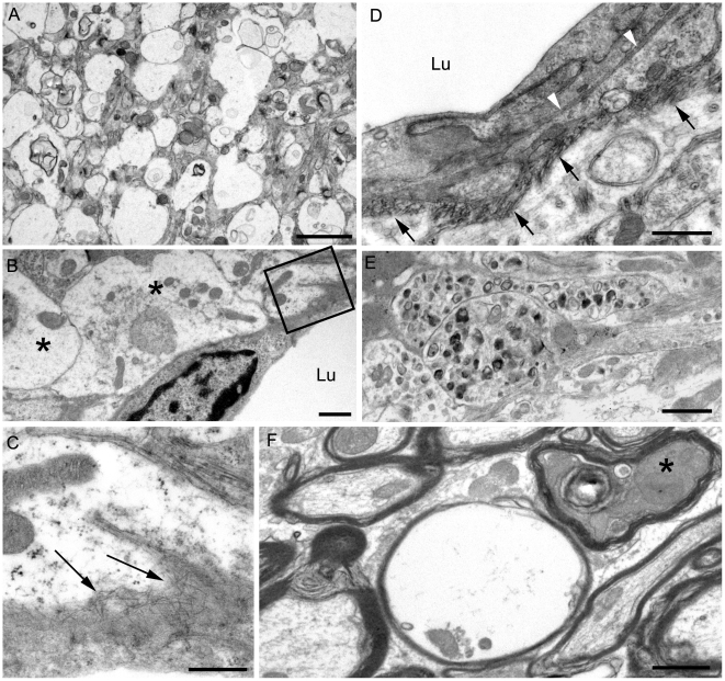
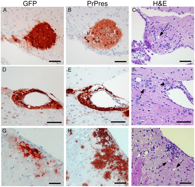
Prion diseases are fatal neurodegenerative diseases of humans and animals characterized by gray matter spongiosis and accumulation of aggregated, misfolded, protease-resistant prion protein (PrPres). PrPres can be deposited in brain in an amyloid-form and/or non-amyloid form, and is derived from host-encoded protease-sensitive PrP (PrPsen), a protein normally anchored to the plasma membrane by glycosylphosphatidylinositol (GPI). Previously, using heterozygous transgenic mice expressing only anchorless PrP, we found that PrP anchoring to the cell membrane was required for typical clinical scrapie. However, in the present experiments, using homozygous transgenic mice expressing two-fold more anchorless PrP, scrapie infection induced a new fatal disease with unique clinical signs and altered neuropathology, compared to non-transgenic mice expressing only anchored PrP. Brain tissue of transgenic mice had high amounts of infectivity, and histopathology showed dense amyloid PrPres plaque deposits without gray matter spongiosis. In contrast, infected non-transgenic mice had diffuse non-amyloid PrPres deposits with significant gray matter spongiosis. Brain graft studies suggested that anchored PrPsen expression was required for gray matter spongiosis during prion infection. Furthermore, electron and light microscopic studies in infected transgenic mice demonstrated several pathogenic processes not seen in typical prion disease, including cerebral amyloid angiopathy and ultrastructural alterations in perivascular neuropil. These findings were similar to certain human familial prion diseases as well as to non-prion human neurodegenerative diseases, such as Alzheimer's disease.


Conflict of interest statement

The authors have declared that no competing interests exist.

Figures

Figure 1
Figure 1. Brain PrPsen expression levels.
C57BL/10 (Prnp+/+), Prnp+/−, expressing normal anchored mouse PrP, and transgenic mice (tg44+/+ and tg44+/−), expressing only anchorless mouse PrP were compared. Immunoblots were done using brain homogenates made as described in methods, and samples were serially diluted two-fold in sample buffer to give the mg brain equivalents shown on the figure. Bands were detected with monoclonal antibody D13. (A) No PNGase digestion. Lanes 1–3: Prnp+/+, Lanes 4–6: Prnp+/−, Lanes 7–9: tg44+/+ and Lanes 10–12: tg44+/−. (B) After PNGase F treatment. Lanes 1–3: Prnp+/−, Lanes 4–5: tg44+/+, Lanes 6–8: Prnp+/− and 9–11: tg44+/+ mice. Lanes 1–5 were from one experiment and 6–11 were from a separate experiment and results shown are for 4 different mice. Lower apparent molecular weight in PrP of transgenic mice is due to lack of the GPI anchor. Prnp+/− mice appeared to have 4-fold more brain PrPsen than tg44+/+ mice (compare lanes 2 and 4, also lanes 7 and 10). Data shown are for tg44+/+ and tg44+/− mice. By immunoblot PrPsen expression levels in tg23 mice were indistinguishable from those in tg44 mice (data not shown).
Figure 2
Figure 2. Survival curves for scrapie-infected mice.
Transgenic mice (tg44+/+ and tg23+/+) expressing only anchorless PrP, C57BL/10 (Prnp+/+) and Prnp+/− mice, expressing normal anchored mouse PrP were compared. Mice were inoculated intracerebrally with 22L scrapie (panel A) and RML scrapie (panel B) and observed weekly for development of disease (see Table 1). Mice were euthanized when clinical signs were severe as described in Table 1. N values for each group are as follows: Panel A (22L): Prnp+/+, 11; Prnp+/−, 17; tg44+/+, 23; tg23+/+, 10. Panel B (RML): Prnp+/+, 8; tg44+/+, 21; tg23+/+, 9.
Figure 3
Figure 3. Detection of PrPres by immunoblot using monoclonal antibody D13.
Comparison of PrPres in brain of 22L scrapie-infected tg44+/+ and tg44+/− mice at the time of clinical disease. All lanes were loaded with 0.25 mg brain tissue equivalents. A clinical Prnp+/− mouse is shown for comparison. Lane 1: Prnp+/− (251 dpi), PrPres bands are seen at 21, 28, and 31 kD; lanes 2–5: tg44+/− mice (567, 589, 594, 594 dpi) and lanes 6–9 tg44+/+ (348, 365, 384, 408 dpi). PrPres bands are at 18 and 23 kD. The sizes are lower in these mice due to lack most carbohydrates and lack of GPI . PrPres levels in tg44+/− mice were approximately 50% lower than in tg44+/+ mice.
Figure 4
Figure 4. Light microscopic histopathology of scrapie-infected C57BL/10 and tg44+/+ transgenic mice.
(A) Whole brain sagittal section of C57BL/10 mouse infected IV with RML scrapie at 180 dpi showing wide distribution of diffuse PrPres stained with monoclonal antibody D13. (B) High power view of C57BL/10 mouse infected IC with RML scrapie at 157 dpi showing diffuse punctuate pattern of PrPres in hippocampus. (C) H&E stain of forebrain of C57BL/10 mouse infected IP with 22L scrapie at 375 dpi. Anterior commissure white matter (WM) is seen in lower right. Numerous scrapie vacuoles (arrows) are visible in surrounding gray matter(GM). (D) Whole brain saggital section of tg44+/+ mouse D554 infected IC with RML scrapie showing dense plaque-like PrPres stained with monoclonal antibody D13 at 341 dpi. PrPres was found in most CNS areas including cerebral cortex, corpus callosum, forebrain, hippocampus, thalamus, hypothalamus, midbrain, colliculi, brainstem, and spinal cord. Cerebellar involvement was minimal after RML infection, as shown in panel 4D, but was strong in cerebellar molecular layer, granular layer and meninges after 22L infection (not shown). (E) Higher power of panel D shows large dense PrP plaques surrounding dentate gyrus of hippocampus often in a perivascular distribution (arrows). Note difference compared to diffuse PrPres staining in panel B. (F) H&E stain of mouse D554 showing vacuoles in the white matter (WM) near the anterior commissure and no vacuoles in surrounding gray matter, i.e. opposite distribution of vacuoles compared to C57BL/10 mouse in panel C. (G) Astrogliosis seen by staining with anti-GFAP in dentate gyrus of mouse D554. (H) H&E stain of dentate gyrus of mouse D554 shows marked neuronal loss in lower arm of gyrus (box and arrow). Boxed outlines region shown in panel I. (I) High power view of lower arm of dentate gyrus outlined in panel H, shows multiple areas of neuronal loss (arrows). Plaques surround this area and one plaque is indicated with the arrowhead at the left. (J) High power view of area shown in panel I shows D13 staining of PrPres plaques impinging on damaged neurons of the dentate gyrus (arrow). Arrowhead shows plaque around blood vessel in upper left corner. (K) Deposition of amyloid precursor protein, APP, (red-brown stain) adjacent to area of neuronal loss (arrows) in dentate gyrus of same area shown in panels I and J. (L) Abnormal axonal proliferation shown by staining with anti-neurofilament protein in area of neuronal loss (arrows) in dentate gyrus of mouse D554. (M) Anti- neurofilament protein staining of dentate gyrus of uninfected control mouse shows no neuronal damage or abnormal axonal staining within the gyrus. Scale bars in panels B, E, G, and H are 100 microns; all other scale bars are 50 microns. Similar pathological changes were seen at the time of clinical disease in both tg44+/+ and tg23+/+ mice infected with either 22L or RML strains of scrapie. D13 staining of PrPres in panels E and J was similar to results described previously in tg44+/− and tg23+/− mice at ≥498 dpi .
Figure 5
Figure 5. Immunological detection of PrPres in brain at both light and electron microscopic levels.
The 22L scrapie-infected anchorless PrP tg44+/+ mouse shown was clinically positive at 377 dpi. (A–B) Light microscopy of 1 µm thick plastic-embedded tissue labelled with monoclonal antibody 1A8. (A) shows intravascular and perivascular PrPres. In (B), the marked vascular amyloid infiltration is associated with occlusion of the vascular lumen (boxes indicate occluded lumens of two vessels). When these vessels were visualised in the electron microscope the smooth muscle of the vascular media was totally replaced by amyloid and an amorphous electron dense material filled the lumen (not shown). (C–F) Electron microscopy. (C) Low power view of large PrPres amyloid plaque adjacent to a small artery. Vessel lumen is in upper left corner and an endothelial cell with a prominent nucleus is to the right of the lumen. Silver enhanced gold-labeled PrPres is seen within the basement membrane (BM) and within the heavy amyloid accumulation which partially replaces the smooth muscle media layer. Amyloid bundles radiate away from the vessel and through the neuropil at the bottom right. (D) A high magnification illustration of (C) showing PrPres labelling on small bundles of amyloid fibrils at the periphery of the plaque. (E) Marked PrPres accumulation at the endothelial and pericyte basement membranes (arrows) and extending into narrow extracellular spces between nearby neurites and perivascular glial processes. Asterix (*) shows area of astrocytic cytoplasmic swelling. Lu; lumen. (F) Neuropil of cerebrum showing immunogold label for PrPres present over the extracellular spaces between neurites bounded by pairs of adjacent plasmalemmae. No visible amyloid fibrils were visible and the spaces between cellular processes were regular and even. This non-fibrillar PrPres labelling which dissects between neurite and glial cell profiles may extend over large area of neuropil as shown in (G). Asterix (*) on left side shows enlarged glial process with loss of normal cytoplasmic organelles. Similar findings were observed in tg44+/+, tg44+/− and tg23+/− mice. Tg23+/+ mice were not examined by electron microscopy. Scale bars: A and B, 20 µm; C and D, 1 µm; E and G, 2 µm; F, 500 nm.
Figure 6
Figure 6. Ultrastructure of cerebral cortex and cerebellum of a 22L scrapie-infected tg44+/+ mouse.
Sections were from same mouse as shown in Figure 5, and were stained with uranyl acetate/lead citrate. (A) Area of neuropil with severe vacuolation where most vacuoles originate within processes and are separated from each other by intact membranes. (B) Several distended astrocytic processes (asterisks) of the perivascular glial limitans are present around a blood vessel Lu: lumen. (C) On higher magnification of boxed area from (B), the endothelial basement is shown to be filled with irregularly orientated amyloid fibrils (arrows). (D) The earliest stage of vascular amyloid is shown. Here the endothelial basement membrane (black arrowheads) is intact, but the pericyte basement membrane (black arrows) is thickened and heavily infiltrated with short amyloid fibrils (white arrowheads). (E) Severe neuritic dystrophy in which several processes show an excessive accumulation of organelles and abnormal electron dense bodies. (F) White matter of the cerebellum showing an empty myelin sheath (seen as vacuoles by light microscopy) and also a dark degenerate axon (asterisk) within an intact myelin sheath. Similar findings were observed in RML scrapie-infected tg44+/+ mice. Scale bars: A, 2 µm; B, E, F, 1 µm; C, 500 nm; D, 500 nm.
Figure 7
Figure 7. Detection of PrPres and vacuolation in brain tissue of PrPnull and tg44+/− mice with C57BL/6 brain grafts.
Grafts expressed green fluorescent protein (GFP), and mice were infected IC with 22L scrapie approximately 5 weeks after grafting. Panels A–C, PrPnull recipient at 261 dpi; Panels D–F, tg44+/− recipient at 261 dpi; Panels G–I, tg44+/− recipient at 200 dpi. Panels A, D, G show staining with anti- GFP which detects constitutive GFP expression in the C57BL/6 donor tissue. Panels B, E, H show D13 staining of PrPres. Panels C, F, I show H&E staining to detect scrapie-induced vacuoles indicated by arrows.

Similar articles

Cited by

References

    1. Aguzzi A, Polymenidou M. Mammalian prion biology: one century of evolving concepts. Cell. 2004;116:313–327. - PubMed
    1. Di Bari MA, Chianini F, Vaccari G, Esposito E, Conte M, et al. The bank vole (Myodes glareolus) as a sensitive bioassay for sheep scrapie. J Gen Virol. 2008;89:2975–2985. - PubMed
    1. Nonno R, Di Bari MA, Cardone F, Vaccari G, Fazzi P, et al. Efficient transmission and characterization of Creutzfeldt-Jakob disease strains in bank voles. PLoS Pathog. 2006;2:e12. doi: 10.1371/journal.ppat.0020012. - DOI - PMC - PubMed
    1. Jeffrey M, Goodsir CM, Bruce ME, McBride PA, Fraser JR. In vivo toxicity of prion protein in murine scrapie: ultrastructural and immunogold studies. Neuropathol Appl Neurobiol. 1997;23:93–101. - PubMed
    1. Gonzalez L, Martin S, Begara-McGorum I, Hunter N, Houston F, et al. Effects of agent strain and host genotype on PrP accumulation in the brain of sheep naturally and experimentally affected with scrapie. J Comp Pathol. 2002;126:17–29. - PubMed

Publication types

MeSH terms