. 2010 Mar 5;6(3):e1000794.
doi: 10.1371/journal.ppat.1000794.

Direct visualization by cryo-EM of the mycobacterial capsular layer: a labile structure containing ESX-1-secreted proteins

Affiliations

Direct visualization by cryo-EM of the mycobacterial capsular layer: a labile structure containing ESX-1-secreted proteins

Musa Sani et al. PLoS Pathog. .

Abstract

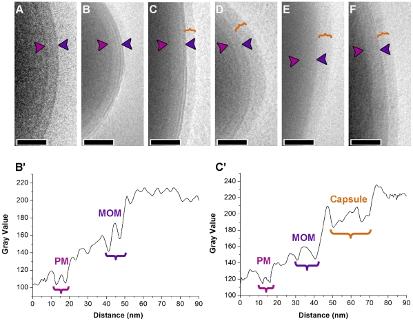
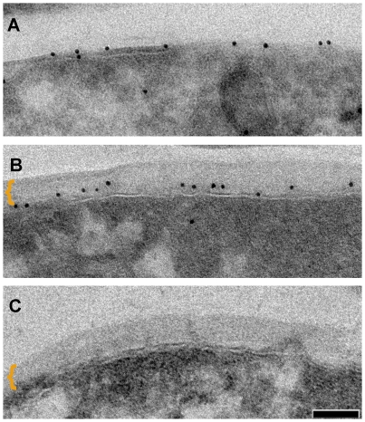
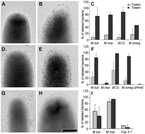
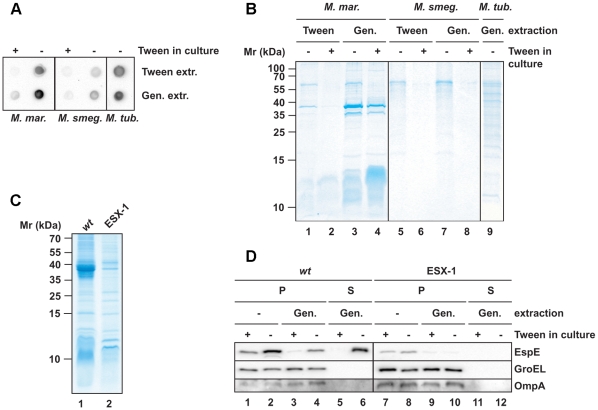
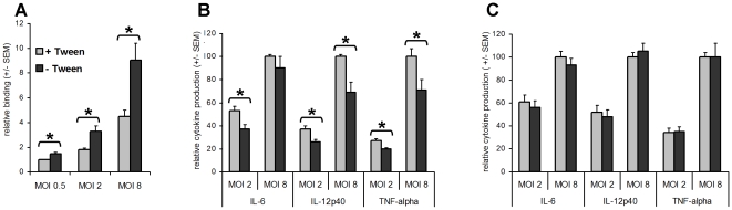
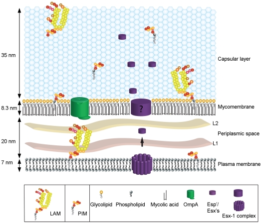
The cell envelope of mycobacteria, a group of Gram positive bacteria, is composed of a plasma membrane and a Gram-negative-like outer membrane containing mycolic acids. In addition, the surface of the mycobacteria is coated with an ill-characterized layer of extractable, non-covalently linked glycans, lipids and proteins, collectively known as the capsule, whose occurrence is a matter of debate. By using plunge freezing cryo-electron microscopy technique, we were able to show that pathogenic mycobacteria produce a thick capsule, only present when the cells were grown under unperturbed conditions and easily removed by mild detergents. This detergent-labile capsule layer contains arabinomannan, alpha-glucan and oligomannosyl-capped glycolipids. Further immunogenic and proteomic analyses revealed that Mycobacterium marinum capsule contains high amounts of proteins that are secreted via the ESX-1 pathway. Finally, cell infection experiments demonstrated the importance of the capsule for binding to cells and dampening of pro-inflammatory cytokine response. Together, these results show a direct visualization of the mycobacterial capsular layer as a labile structure that contains ESX-1-secreted proteins.


Conflict of interest statement

The authors have declared that no competing interests exist.

Figures

Figure 1
Figure 1. Visualization of the capsule in its native state.
Cryo electron micrographs of intact S. flexneri cell plunge frozen (A) depicts the typical cell envelope profile using this method of sample preparation. (B) M. smegmatis cells grown with both chemical and mechanical perturbation shows a cell envelope with morphology similar to S. flexneri. Similar cells cultured in an unperturbed state before freezing (C) shows the presence of an extra layer (bracket) surrounding the mycomembrane. Corresponding layers are also observed surrounding the mycomembranes of M. tuberculosis (D), M. marinum (E), and M. bovis BCG (F). The density profiles in B' and C' were obtained from images corresponding to B and C respectively. Images were averaged over a width of 75 pixels. The outermost layer varies in thickness from negligible to considerable (40 nm; see Table S1) indicating the unstable nature of this layer. Arrow heads point to plasma membrane (PM; magenta) and outer membrane/mycomembrane (MOM; blue). Scale bars: 100 nm.
Figure 2
Figure 2. Distinction between the mycomembrane and the outermost layer.
Immuno-labeling of outer membrane porin OmpATb on thin sections of M. smegmatis overexpressing OmpATb (mechanically agitated and detergent treated culture) (A) and non-perturbed culture (B) as compared to M. smegmatis wt (C). Outermost layer indicated by bracket. Scale bar: 50 nm.
Figure 3
Figure 3. Effect of detergent on the localization of capsular components and the detection of ESX-1 proteins.
M. smegmatis (A and B), M. tuberculosis (D and E) and M. marinum (G and H) were grown under both perturbed (A,D and G) and unperturbed conditions (B,E and H), fixed and probed with anti-α-glucan (A–B) and anti PIM/capLAM (D–E) to demonstrate that this extra layer is of capsular origin. (G–H) demonstrates the presence of anti-EspE on bacterial surface/capsule. Histogram (C,F and I) respectively displays the variations in anti glucan, anti PIM/capLAM and anti-EspE labeling of bacteria due to change in the culturing conditions using M. smegmatis ΔPimE k.o. and M. marinum fas 3.1 eccCb1 mutant as controls. The ordinate (defined as percentage of bacteria with ≥10 gold particles) represents the average ± standard error of labeled cells from 3 independent experiments. Scale bars: 250 nm.
Figure 4
Figure 4. Extraction of mycobacterial capsules by mild detergent.
Cell surface extracts from mycobacteria that were cultured under perturbed and unperturbed conditions (first normalized to proteins amounts of the pellet fraction) were prepared by incubation with 1% Tween-80 or 0.25% Genapol X-080. (A and B) Extracted fractions were analyzed for the presence of α-glucan by a spot blot assay (A) and for the protein content by SDS-PAGE and Coomassie staining (B). (C) Comparisons of the extracted proteins from wt M. marinum and an ESX-1 mutant by SDS-PAGE and Coomassie staining. (D) Immunoblot showing the Genapol-extractability of EspE. Non-extracted material (P) was separated from the extracted material (S) by centrifugation and equivalent amounts were loaded. As negative controls, the mycomembrane protein OmpA and the cytosolic protein GroEL were analyzed.
Figure 5
Figure 5. Capsular layer promotes binding to macrophages and inhibits the pro-inflammatory cytokine response.
The results represent the mean relative binding ± standard error of the mean (SEM) of the pooled data from three independent donors. (A) The percentage of macrophages associated with Tween. M. bovis BCG expressing dsRed bacteria was determined using flow cytometry. Binding of Tween-treated M. bovis BCG at MOI 0.5 (1.13% +/− 0.24 (SD)) is set at 1. (B and C) Macrophages were stimulated with/without Tween treatment M. bovis BCG (B) or M. smegmatis (C) at MOIs 2 and 8. Cells stimulated with PBS served as a control. After 24 h, the supernatants were harvested and the amount of IL-12p40, IL-6, and TNF-α was determined using ELISA. The results represent the mean relative cytokine production (± SEM) of the pooled data from at least three independent donors. Cytokine production following stimulation with Tween-treated bacteria at MOI 8 is set at 100 [for M. bovis BCG, IL-6: 12087±3056, IL-12p40: 2928±618, TNFα: 38490±6446; for M. smegmatis, IL-6: 61629±17411, IL-12p40: 22693±2100, TNFα: 122663±17350 (all in pg mL−1 ± SEM)]. An asterisk indicates significant differences (p<0.05).
Figure 6
Figure 6. The spatial organization of the mycobacterial cell envelope exhibiting the capsule.
This scheme represents the relative size and organization of the different layers of the envelope including the plasma membrane, mycomembrane, periplasmic space and capsular layer. The positions of some of the constituents analyzed are depicted.

References

    1. Gagliardi MC, Lemassu A, Teloni R, Mariotti S, Sargentini V, et al. Cell wall-associated alpha-glucan is instrumental for Mycobacterium tuberculosis to block CD1 molecule expression and disable the function of dendritic cell derived from infected monocyte. Cell Microbiol. 2007;9:2081–2092. - PubMed
    1. Daffe M, Etienne G. The capsule of Mycobacterium tuberculosis and its implications for pathogenicity. Tubercle Lung Dis. 1999;79:153–169. - PubMed
    1. Minnikin DE. Lipids: complex lipids, their chemistry, biosynthesis and roles.The Biology of Mycobacteria. In: Ratledge C, Standford J, editors. The Biology of Mycobacteria. London: Academic; 1982. pp. 95–184.
    1. Brennan PJ, Crick DC. The cell-wall core of Mycobacterium tuberculosis in the context of drug discovery. Curr Top Med Chem. 2007;7:475–488. - PubMed
    1. Hoffmann C, Leis A, Niederweis M, Plitzko JM, Engelhardt H. Disclosure of the mycobacterial outer membrane: cryo-electron tomography and vitreous sections reveal the lipid bilayer structure. Proc Natl Acad Sci U S A. 2008;105:3963–3967. - PMC - PubMed

Publication types

MeSH terms

Substances