. 2010 Mar 5;6(3):e1000701.
doi: 10.1371/journal.ppat.1000701.

Direct restriction of virus release and incorporation of the interferon-induced protein BST-2 into HIV-1 particles

Affiliations

Direct restriction of virus release and incorporation of the interferon-induced protein BST-2 into HIV-1 particles

Kathleen Fitzpatrick et al. PLoS Pathog. .

Abstract

Investigation of the Vpu protein of HIV-1 recently uncovered a novel aspect of the mammalian innate response to enveloped viruses: retention of progeny virions on the surface of infected cells by the interferon-induced, transmembrane and GPI-anchored protein BST-2 (CD317; tetherin). BST-2 inhibits diverse families of enveloped viruses, but how it restricts viral release is unclear. Here, immuno-electron microscopic data indicate that BST-2 is positioned to directly retain nascent HIV virions on the plasma membrane of infected cells and is incorporated into virions. Virion-incorporation was confirmed by capture of infectivity using antibody to the ectodomain of BST-2. Consistent with a direct tethering mechanism, we confirmed that proteolysis releases restricted virions and further show that this removed the ectodomain of BST-2 from the cell surface. Unexpectedly, enzymatic cleavage of GPI anchors did not release restricted virions, weighing against models in which individual BST-2 molecules span the virion and host cell membranes. Although the exact molecular topology of restriction remains unsolved, we suggest that the incorporation of BST-2 into viral envelopes underlies its broad restrictive activity, whereas its relative exclusion from virions and sites of viral assembly by proteins such as HIV-1 Vpu may provide viral antagonism of restriction.


Conflict of interest statement

The authors have declared that no competing interests exist.

Figures

Figure 1
Figure 1. Punctate cell surface distribution of BST-2 and surface down-regulation by HIV-1 encoding the Vpu protein.
HeLa cells were left untransfected (A) or transfected with a plasmid expressing the complete wild type HIV-1 genome (B) or with a plasmid expressing an HIV-1 genome lacking vpu (C). The next day, the cells were fixed and stained without permeabilization using an antibody to the BST-2 ectodomain and a secondary labeling system including streptavidin-conjugated cadmium selenide/zinc sulfide nanocrystals (quantum dots; Qdot 625 nm). Nuclei were stained with DAPI. Data were acquired as a Z-series of images using an Olympus laser scanning confocal microscope. The images shown are projections of the entire Z-series for each field. (D) HeLa cells (untransfected) were plated on cover glasses, then fixed in formaldehyde and stained for surface BST-2 exactly as described above using Qdot 625 nanocrystals. After staining, the cells were further fixed in glutaraldehyde and osmium tetroxide, followed by conventional embedding and processing for thin-section transmission electron microscopy. Images were obtained using a JEOL 1200C microscope operated at 80 keV.
Figure 2
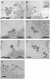
Figure 2. Electron microscopic evidence of direct virion tethering and virion-incorporation of BST-2.
HeLa cells were transfected to express the complete HIV-1 genome (wild type; A, C, and E) or a genome lacking vpu (Δvpu; B, D, F, and G), then stained for cell surface BST-2 and processed for electron microscopy as described in the legend of Figure 1. Panels A-F are images of thin sections obtained using a JEOL 1200C microscope operated at 80 keV. Panel G is a projection view of a thick section (approximately 250 nm) derived from a tomographic tilt series of images obtained using an FEI Titan electron microscope operated at 300 keV.
Figure 3
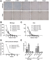
Figure 3. Capture of infectious HIV-1 virions using antibody to BST-2.
A. HIV-1 virions were produced by transfection of HeLa cells, which express endogenous BST-2, either with a plasmid expressing the wild type HIV-1 genome or a genome lacking vpu (Δvpu). Forty-eight hours later, cell-free culture supernatants were collected, incubated with antibody to CD44, BST-2, CD45, or an IgG2a isotype control, then captured using magnetic microbeads coated with staphylococcal protein G. Immuno-captured virions were resuspended in the original culture volumes. Virions, uncaptured or captured using the indicated antibodies, were used to infect CD4-positive HeLa cells containing an LTR-β-galactosidase indicator construct. Two days later, infectious centers were visualized by incubation with X-gal to generate blue colored foci. Images of individual wells were acquired using a CCD camera. The wells shown are the result of infection using undiluted preparations, which were each equivalent to 200 µl of the original, uncaptured cell-free culture supernatants. The uncaptured preparation of wild type virus contained five-fold more HIV-1 p24 capsid antigen than that of Δvpu, consistent with the restriction of virion-release by endogenous BST-2 and counteraction by Vpu (data not shown). B and C. An independently prepared set of virions was produced from HeLa cells, subjected to immuno-capture, and infectivity was measured as described for panel A. Each preparation was serially diluted as shown (“0” indicates undiluted sample) and used to infect CD4-positive HeLa cells in duplicate. Infectious centers were counted using image analysis software; error bars are the actual values of duplicate wells and in some cases are too small to see. The data shown are representation of three independent experiments. D. Virions (wild type) were produced by transfection of HEK293T cells lacking endogenous BST-2, subjected to immuno-capture using antibody to BST-2 or an antibody isotype control, and infectivity was measured as in panels B and C. Data are the averages of duplicate wells; the error bars are too small to see. E. The fraction of the total viral p24 capsid antigen that was recovered in these immuno-capture experiments was measured using an ELISA. Error bars are the standard deviation of values obtained from three capture experiments using independent preparations of viruses made from HeLa cells.
Figure 4
Figure 4. Virion-associated BST-2 detected by immunoblot.
HIV-1 virions were produced by transfection of HeLa cells, which express endogenous BST-2, either with a plasmid expressing the wild type HIV-1 genome or a genome lacking vpu. Twenty-four hours after transfection, culture supernatants were centrifuged at 800 g for 8 minutes to remove cellular debris. Virions were concentrated by centrifugation of the clarified supernatants at 23,000 g for three hours, and then resuspended in SDS- and DTT-containing buffer for quantitation of p24 capsid by ELISA and for analysis of BST-2 and p24 content by western blot. A. Blots were probed for BST-2. Left lanes were normalized by volume: the wild type sample contained 85 ng of p24 antigen, whereas the Δvpu sample contained 4 ng of p24 antigen. The greater p24 content of the wild type preparation reflects the antagonism of BST-2 mediated restriction of virion-release by Vpu. Right lanes were normalized by p24 antigen content (5 ng for each sample as determined by ELISA). B. Samples analyzed in A were reanalyzed including a dilution series of wild type virions and detection of p24 antigen by immunoblot. Stars indicate lanes with approximately equal content of p24. Western blots for BST-2 were performed as previously described . Molecular weights are in kilodaltons.
Figure 5
Figure 5. Release of nascent virions and the BST-2 ectodomain by proteolysis of the cell surface with subtilisin; models of direct tethering.
A. HeLa cells were transfected to express the complete HIV-1 genome (WT) or a genome lacking vpu (ΔVpu). The next day, culture supernatants were removed to measure the amount of spontaneously released virions (“culture sup”). The cells were then incubated with buffer (control) or buffer containing 1 mg/ml subtilisin for 15 minutes at 37C. Proteolysis was stopped by the addition of PMSF to 5 mM; the cells were pelleted, and the supernatants were removed to measure the amount of virions released (“subtilisin sup”). The cells were then directly lysed into Triton-X 100 containing buffer to measure cell-associated viral antigen. The concentrations of HIV-1 p24 capsid antigen were measured in all samples by ELISA. “Total” indicates the total p24 antigen produced by the culture, i.e., the sum of that released spontaneously, after incubation with buffer or subtilisin, and cell-associated. “Cellular total” indicates the amount of p24 antigen initially associated with the cells, i.e., the sum of that released by buffer or subtilisin and cell-associated. “Culture+subtilisin sup” indicates the total amount of p24 antigen releasable from the cell surface, i.e., the sum of that released spontaneously and after incubation with buffer or subtilisin. Data are the percentages of p24 released; error bars indicate the values of duplicate measurements. B. HeLa cells were treated with buffer or subtilisin as above; proteolysis was quenched as above; and the cells were stained with antibody to the BST-2 ectodomain or with an isotype control antibody, and then analyzed by flow cytometry. Lightly shaded curve, antibody isotype control; unshaded curve, BST-2 after proteolysis of the cell surface using subtilisin; darkly shaded curve, BST-2 on untreated cells. C. Soluble BST-2 ectodomain (residues 49–180 as described previously [6]) was incubated with the indicated amounts of subtilisin for fifteen minutes before analysis by SDS-PAGE and detection by immunoblot with antibody specific to the BST-2 ectodomain. D. Model of ectodomain interaction. E. Model of virion-cell membrane spanning dimer: anti-parallel orientation. F. Model of virion-cell membrane spanning dimer: parallel orientation.
Figure 6
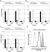
Figure 6. Persistence of virion tethering despite cleavage with PI-specific PLC either with or without disulfide reduction.
Experiments were performed as described in the legend of Figure 5, except that rather than subtilisin, specific treatments were used to test models of tethering. A. Release data using bacterial phosphatidyl inositol-specific phosphoplipase C (PI-PLC; 2.5 U/ml). B. Release data using 5 mM dithiothreitol (DTT) at 37C. C. Release data using PI-PLC followed by DTT. In panels A-C, data are the percentages of p24 released; error bars indicate the values of duplicate measurements. D. Confirmation of PI-PLC activity using CD55 (decay accelerating factor) as a control protein. HeLa cells were stained with either a FITC-conjugated isotype control antibody (dotted line), or a FITC conjugated specific antibody to CD55 (dark-outlined open curve and shaded curve), before analysis by flow cytometry. Cells treated with PI-PLC (2.5 U/ml) are indicated by the dark-outlined open curve, whereas control cells incubated in buffer only are indicated by the shaded curve.

Similar articles

Cited by

References

    1. Malim MH, Emerman M. HIV-1 accessory proteins–ensuring viral survival in a hostile environment. Cell Host Microbe. 2008;3:388–398. - PubMed
    1. Klimkait T, Strebel K, Hoggan MD, Martin MA, Orenstein JM. The human immunodeficiency virus type 1-specific protein vpu is required for efficient virus maturation and release. J Virol. 1990;64(2):621–629. - PMC - PubMed
    1. Varthakavi V, Smith RM, Bour SP, Strebel K, Spearman P. Viral protein U counteracts a human host cell restriction that inhibits HIV-1 particle production. Proc Natl Acad Sci U S A. 2003;100:15154–15159. - PMC - PubMed
    1. Neil SJ, Zang T, Bieniasz PD. Tetherin inhibits retrovirus release and is antagonized by HIV-1 Vpu. Nature. 2008;451:425–430. - PubMed
    1. Van Damme N, Goff D, Katsura C, Jorgenson RL, Mitchell R, et al. The interferon-induced protein BST-2 restricts HIV-1 release and is downregulated from the cell surface by the viral Vpu protein. Cell Host Microbe. 2008;3:245–252. - PMC - PubMed

Publication types

MeSH terms