. 2010 Mar 28;39(12):2964-71.
doi: 10.1039/b922555b. Epub 2010 Jan 11.

Dual functions of NifEN: insights into the evolution and mechanism of nitrogenase

Affiliations

Dual functions of NifEN: insights into the evolution and mechanism of nitrogenase

Yilin Hu et al. Dalton Trans. .

Abstract

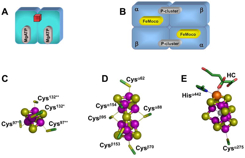
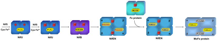
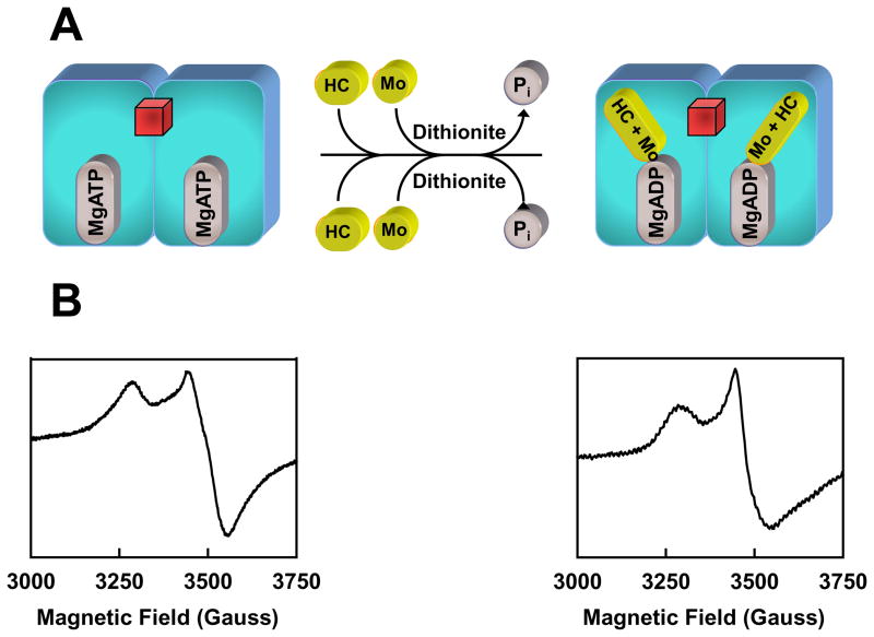
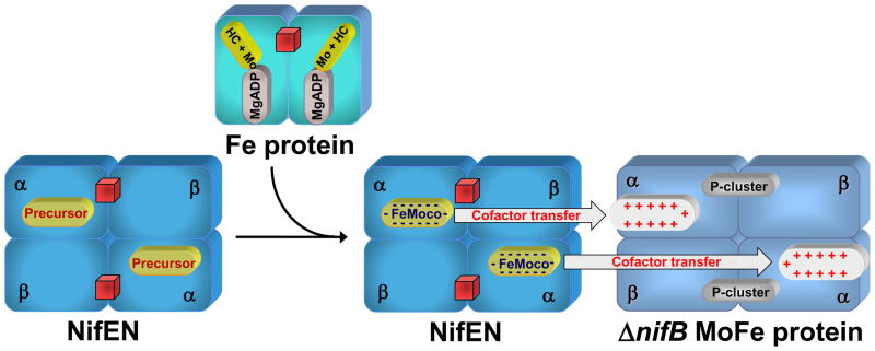
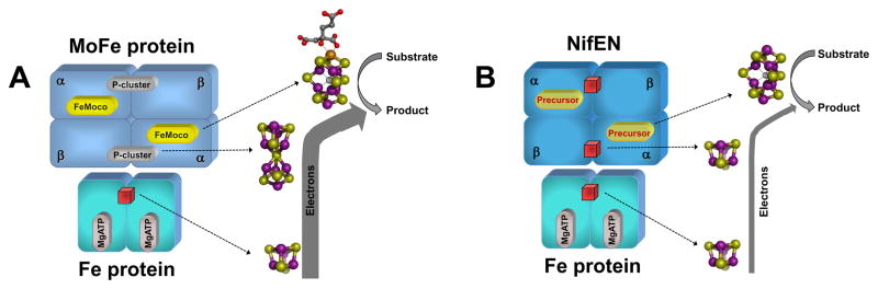
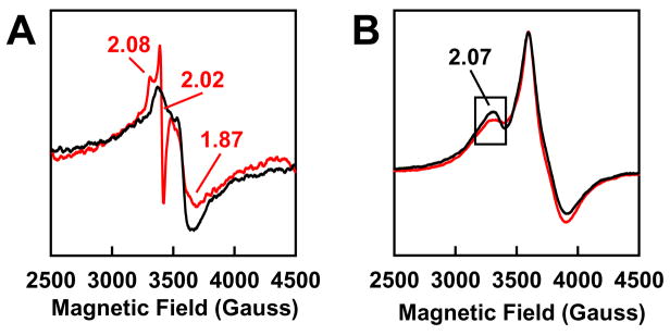
Nitrogenase catalyzes the nucleotide-dependent conversion of dinitrogen to ammonia at the iron-molybdenum cofactor (FeMoco) center of its molybdenum-iron (MoFe) protein component. Biosynthesis of FeMoco is arguably one of the most complex processes in the field of bioinorganic chemistry, which involves the participation of a number of nif (nitrogen fixing) gene products. One key player in this process, NifEN (encoded by nifE and nifN), is homologous to the MoFe protein with regard to both the primary sequences and the types of the metal centers. Recently, an all-iron precursor has been identified on NifEN, which closely resembles the Fe/S core structure of the mature cofactor. Such a precursor-bound form of NifEN has not only served as an excellent platform for the investigation of FeMoco assembly, but also facilitated the examination of the capacity of NifEN as a catalytic homolog of MoFe protein. This perspective will focus on the recent advances toward elucidating the dual functions of NifEN in nitrogenase assembly and catalysis, and the insights afforded by these advances into the evolution and mechanism of nitrogenase.


Figures

Fig. 1
Fig. 1. Component proteins and metal centers of Mo-nitrogenase
(A,B) Schematic presentations of the α2-dimeric Fe protein (A), which contains a [Fe4S4] cluster (red cube) at the subunit interface and a MgATP binding site within each subunit; and the α2β2-tetrameric MoFe protein (B), which contains a pair of unique clusters in each αβ-subunit dimer: the P-cluster ([Fe8S7]) at the αβ-subunit interface; and the FeMoco ([MoFe7S9X], where X = C, N, or O) within the α-subunit. (C-E) Structures of the metal centers in Fe protein and MoFe protein. In the Fe protein, the [Fe4S4] cluster is ligated by Cys97 and Cys132 from both subunits (C). In the MoFe protein, the P-cluster is ligated by three Cys residues from the α-subunit (Cysα62, Cysα88 and Cysα154) and three Cys residues from the β-subunit (Cysβ70, Cysβ95 and Cysβ153) (D); and the FeMoco is ligated by only two ligands: Cysα275 and Hisα442. The atoms of the metal centers are colored as follows: Mo, orange; Fe, purple; S, yellow; O, red; C, green; X (C, N or O), light gray. HC stands for homocitrate. These presentations are generated in PYMOL using 1N2C and 1M1N PDB coordinates.,
Fig. 2
Fig. 2. Biosynthetic pathway of FeMoco
NifS and NifU launch the process by synthesizing small Fe/S fragments, such as the [Fe2S2] clusters (red diamonds) and the [Fe4S4] clusters (red cubes). These small Fe/S building blocks are assembled into a large Fe/S core on NifB and further processed on NifEN with the assistance of Fe protein. Upon the completion of assembly on NifEN, the mature FeMoco is subsequently delivered to its target location within the MoFe protein, resulting in the formation of an active holo-MoFe protein. The [Fe4S4] clusters in Fe protein and NifEN are represented by red cubes.
Fig. 3
Fig. 3. Comparison between the primary sequences of NifEN and MoFe protein
(A) Partial sequence alignment between the α-subunits of MoFe protein (NifD) and NifEN (NifE) of Azotobacter vinelandii. (B) Partial sequence alignment between the β-subunits of MoFe protein (NifK) and NifEN (NifN) of A. vinelandii. Four of the six P-cluster ligands in MoFe protein, Cysα62, Cysα88, Cysα154 and Cysβ95, are preserved as Cysα37, Cysα62, Cysα124 and Cysβ81, respectively, in NifEN (A, B). One of the two FeMoco ligands in MoFe protein, Cysα275, is preserved as Cysα250 in NifEN; whereas the other ligand, Hisα442, is replaced by Asnα418 (A). The homocitrate anchor in MoFe protein, Lysα426, is replaced by a homologous residue, Argα402, in NifEN. Two MoFe protein residues that are important for N2 reduction, Glnα191 and Hisα195, are replaced by Lysα160 and Asnα164, respectively, in NifEN (A). Hisα451 and Trpα444, which could secure FeMoco at the cofactor binding site of MoFe protein, are replaced by Alaα426 and Gluα420, respectively, in NifEN (A). Argα97 and Lysα315, which are part of the positively charged insertion funnel in MoFe protein, are replaced by Glyα71 and Leuα290, respectively, in NifEN (A).
Fig. 4
Fig. 4. Precursor- and FeMoco-bound forms of NifEN
(A, C) Dithionite-reduced (upper traces) and indigo disulfonate (IDS)-oxidized (lower traces) EPR spectra of precursor-(A) and FeMoco- (C) bound NifEN. The conversion of precursor (A) to FeMoco (C) is accompanied by the appearance of a FeMoco-like, S = 3/2 signal in the dithionite-reduced spectrum (C, upper trace) and the current disappearance of the g = 1.92 feature in the IDS-oxidized spectrum (C, lower trace). The g values are indicated. (B, D) XAS/EXAFS-derived structures of the NifEN-associated precursor (B) and FeMoco (D). Both structures resemble that of the mature cofactor, except for the absence of Mo and homocitrate from the NifEN-associated precursor (B) and an asymmetric coordination of Mo in the NifEN-associated FeMoco (C). The atoms of the metal centers are colored as follows: Mo, orange; Fe, purple; S, yellow; O, red; C, dark gray; X (C, N or O), light gray. These presentations are generated in PYMOL using 1N2C and 1M1N PDB coordinates.,
Fig. 5
Fig. 5. Mobilization of Mo and homocitrate by Fe protein
(A) In the presence of reductant, Mo and homocitrate (HC) can be “loaded” onto the Fe protein upon ATP hydrolysis. Mo may enter the Fe protein by attaching to the position that corresponds to the γ-phosphate of ATP following the hydrolysis of ATP. Subsequently, the loaded Fe protein can deliver Mo and HC to the NifEN-associated precursor and transform the precursor into a fully matured FeMoco. The [Fe4S4] cluster of the Fe protein is represented by a red cube. (B) EPR spectra of the MgATP-bound Fe protein (left) and the Fe protein loaded with Mo and HC upon ATP hydrolysis (right).
Fig. 6
Fig. 6. Maturation and incorporation of FeMoco
Fe protein loaded with Mo and homocitrate (HC) can serve as a direct Mo/HC source for the maturation of NifEN-bound precursor. Incorporation of Mo and HC into the precursor completes the FeMoco maturation on NifEN. Subsequently, the fully matured FeMoco is transferred from NifEN to its target location within the MoFe protein upon direct protein-protein interactions. Charge-based interactions likely play an important role in the transfer of FeMoco between NifEN and MoFe protein, during which process the negatively charged FeMoco is inserted into the MoFe protein via a positively charged insertion funnel. The [Fe4S4] clusters of Fe protein and NifEN are represented by red cubes.
Fig. 7
Fig. 7. Components of the electron transfer chains in nitrogenase and its homolog
(A) In nitrogenase (i.e., Fe protein/MoFe protein), electrons likely flow from the [Fe4S4] cluster of the Fe protein to the P-cluster and then the FeMoco of the MoFe protein, where substrate reduction occurs. (B) In nitrogenase homolog (i.e., Fe protein/NifEN), electrons could flow from the [Fe4S4] cluster of the Fe protein to the [Fe4S4] cluster and then the all-Fe FeMoco homolog (or precursor) of NifEN. However, there is a much reduced electron flux through the Fe protein/NifEN system (indicated by a thinner arrow), which is likely due to the limited electron transfer capacity of the [Fe4S4] cluster at the “P- cluster site” of NifEN (B). The [Fe4S4] clusters in Fe protein and NifEN are represented by red cubes. The atoms of the metal centers are colored as follows: Mo, orange; Fe, purple; S, yellow; O, red; C, dark gray; X (C, N or O), light gray. These presentations are generated in PYMOL using 1N2C and 1M1N PDB coordinates.,
Fig. 8
Fig. 8. EPR features of NifEN upon substrate turnover
EPR spectra of NifEN under turnover (red) and non-turnover (black) conditions of C2H2 (A) and N3 (B). Turnover samples contained MgATP (red), which is absent form non-turnover samples (black). Spectra were recorded at 30 K (A) and 6 K (B), respectively. The g values are indicated.

Similar articles

Cited by

References

    1. Burgess BK, Lowe DJ. Chem Rev. 1996;96:2983–3012. - PubMed
    1. Howard JB, Rees DC. Chem Rev. 1996;96:2965–2982. - PubMed
    1. Smith BE. Adv Inorg Chem. 1999;47:159–218.
    1. Rees DC, Tezcan FA, Haynes CA, Walton MY, Andrade SL, Einsle O, Howard JB. Philo Trans R Soc London. 2005;A363:971–984. - PubMed
    1. Howard JB, Rees DC. Proc Natl Acad Sci USA. 2006;103:17088–17093. - PMC - PubMed

Publication types

MeSH terms