. 2010 Jul;59(7):1083-95.
doi: 10.1007/s00262-010-0835-z. Epub 2010 Mar 10.

Induction of tumor-specific acquired immunity against already established tumors by selective stimulation of innate DEC-205(+) dendritic cells

Affiliations

Induction of tumor-specific acquired immunity against already established tumors by selective stimulation of innate DEC-205(+) dendritic cells

Keiichi Moriya et al. Cancer Immunol Immunother. 2010 Jul.

Abstract

Two major distinct subsets of dendritic cells (DCs) are arranged to regulate our immune responses in vivo; 33D1(+) and DEC-205(+) DCs. Using anti-33D1-specific monoclonal antibody, 33D1(+) DCs were successfully depleted from C57BL/6 mice. When 33D1(+) DC-depleted mice were stimulated with LPS, serum IL-12, but not IL-10 secretion that may be mediated by the remaining DEC-205(+) DCs was markedly enhanced, which may induce Th1 dominancy upon TLR signaling. The 33D1(+) DC-depleted mice, implanted with syngeneic Hepa1-6 hepatoma or B16-F10 melanoma cells into the dermis, showed apparent inhibition of already established tumor growth in vivo when they were subcutaneously (sc) injected once or twice with LPS after tumor implantation. Moreover, the development of lung metastasis of B16-F10 melanoma cells injected intravenously was also suppressed when 33D1(+) DC-deleted mice were stimulated twice with LPS in a similar manner, in which the actual cell number of NK1.1(+)CD3(-) NK cells in lung tissues was markedly increased. Furthermore, intraperitoneal (ip) administration of a very small amount of melphalan (L: -phenylalanine mustard; L: -PAM) (0.25 mg/kg) in LPS-stimulated 33D1(+) DC-deleted mice helped to induce H-2K(b)-restricted epitope-specific CD8(+) cytotoxic T lymphocytes (CTLs) among tumor-infiltrating lymphocytes against already established syngeneic E.G7-OVA lymphoma. These findings indicate the importance and effectiveness of selective targeting of a specific subset of DCs, such as DEC-205(+) DCs alone or with a very small amount of anticancer drugs to activate both CD8(+) CTLs and NK effectors without externally added tumor antigen stimulation in vivo and provide a new direction for tumor immunotherapy.


Figures

Fig. 1
Fig. 1
a Distribution of 33D1- or DEC-205+ DCs in the spleen as well as in the intestinal epithelium of normal and anti-33D1 mAb injected C57BL/6 mice. To determine the distribution of 33D1- or DEC-205+ DCs, normal C57BL/6 mice were killed and analyzed by flow cytometry. Distributions of the indicated DC-subtypes either in the spleen (SP) (leftsix panels) or in the intestinal epithelium (IE) (rightsix panels) are shown. Among these panels, the uppersix panels were obtained from untreated and the lowersix panels were from anti-33D1 mAb ip injected C57BL/6 mice. b Kinetics for in vivo depletion of 33D1+ DCs by ip injection of anti-33D1 mAb. To determine the continuation of the effect by anti-33D1 mAb treatment, C57BL/6 mice were killed and the percentage of remaining 33D1+ DCs either in the SP (uppersix panels) or in the IE (lowersix panels) was measured by flow cytometry every week for three successive weeks after anti-33D1 mAb ip injection. To achieve complete elimination of 33D1+ DCs, single additional ip injection of anti-33D1 mAb was performed 2 weeks after the initial administration, and their effect was examined for at least another 2 weeks (right four panels)
Fig. 2
Fig. 2
Significant enhancement of IL-12 secretion in 33D1+ DC-depleted mice. To examine the ability to secrete cytokines in the sera of 33D1+ DC-depleted mice (filled bars), they were sc administrated once either with 100 μg LPS, the ligand for TLR4, or with 100 μg poly(I:C), the ligand for TLR3 and the amount of two representative cytokines, IL-10 and IL-12, was measured by ELISA at 2, 6 and 8 h after stimulation. Upper panels show the amounts of IL-12 and lower panels IL-10 after single sc injection of LPS (left two panels) or poly(I:C) (right two panels). Open bars indicate the sera from control mice injected with isotype-matched mAb (rat IgG). Asterisk (*) indicates statistically significant differences (P < 0.05) between 33D1+ DC-depleted and control rat-IgG-treated groups
Fig. 3
Fig. 3
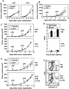
Suppression of tumor growth implanted into the dermis of 33D1+ DC-depleted mice with LPS stimulation and effect of blocking on IL-12 secretion and depletion of CD8+ T cells for suppression. a× 106 syngeneic Hepa1-6 hepatoma cells were injected into the dermis of 33D1+ DC-depleted (filled circle) or control isotype-matched rat-IgG-treated (open circle) C57BL/6 mice. Based on the findings in Fig. 2, 100 µg LPS was injected sc together with the implantation of Hepa1-6 cells (right panel). b, c, e 1 × 106 syngeneic B16-F10 melanoma cells were injected into the dermis of 33D1+ DC-depleted (closed circle) or control rat-IgG-treated (open circle) C57BL/6 mice, which were injected twice with LPS at weekly intervals, 100 μg on day 3 and 50 μg on day 10 after tumor implantation. Asterisk (*) indicates statistically significant differences (P < 0.05) between 33D1+ DC-depleted and control rat-IgG-treated groups on the same day. Mice were also treated with anti-mouse IL-12-specific Ab or control goat IgG just before LPS injection on day 3 and 10 for in vivo neutralization of IL-12 (c) or anti-mouse CD8 Ab (3.155) or control rat IgM at days 7, 6 and 5 before and day 7 after tumor injection for in vivo depletion of CD8+ T cells (e). d Sera were collected from 33D1+ DC-depleted (filled bars) or control mice (open bars) 2 and 6 h after LPS stimulation following ip injection with anti-mouse IL-12 Ab or goat IgG, and IL-12 p40 in the sera was measured by ELISA. f CD4+ and CD8+ lymphocytes in the spleen from mice 7 days after injection with anti-CD8 Ab or rat IgM were analyzed by flow cytometry
Fig. 4
Fig. 4
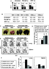
Suppression of lung metastasis of B16-F10 melanoma in 33D1+ DC-depleted mice stimulated with LPS. 33D1+ DC-depleted C57BL/6 mice were injected iv with 1 × 105 B16-F10 melanoma cells via the tail vein per mouse and then stimulated twice with LPS at weekly intervals, 100 µg on day 3 and 50 µg on day 10 after syngeneic tumor injection. Fourteen days after iv injection of B16-F10 melanoma, the number of lung metastases in each mouse was counted either visually (a) or numerically (b). Total volume of the lung in 33D1+ DC-depleted, LPS-treated mice was significantly reduced (P < 0.01) (c)
Fig. 5
Fig. 5
Effect of number of NK cells on lung metastasis development. a, b To determine the cells involved in the suppression of tumor metastasis, lymphocyte subsets obtained from lung tissues with B16-F10 metastasis were analyzed by flow cytometry. The percentage of NK1.1+CD3- NK cells in the lung tissues of untreated mice and LPS-stimulated 33D1+ DC-deleted mice was determined by flow cytometry analysis (a). The results are shown as the mean of the percentage ± SEM from 3 to 4 mice (b). Asterisk (*) indicates statistical differences (P = 0.058) between the 33D1+ DC-depleted group and untreated group. cf Effect of depletion of NK cells in vivo by anti-asialo GM1-specific Ab. NK cell-depleted mice were injected iv with 1 × 105 B16-F10 melanoma cells and stimulated twice with LPS at weekly intervals, 100 µg on day 3 and 50 µg on day 10 after tumor injection. Fourteen days after iv injection of B16-F10 melanoma, lung (c) and SP were removed from the mice and the weight of each lung was measured (d). Asterisk (*) indicates statistically significant differences (P < 0.05) between the control group treated with both rabbit serum with rat IgG and each treated group. Percentages of NK1.1+CD3 cells (e) and NK activity (f) in the lung and SP cells were analyzed by flow cytometry and 51Cr-release assay using YAC-1 cells as targets. The results are shown as the mean ± SEM of triplicates from pooled lung or SP cells obtained from 3 to 4 mice. The effector: target (E:T) ratio is 100:1 or 80:1 in spleen or lung cells, respectively (f)
Fig. 6
Fig. 6
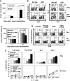
Suppression of B16-OVA tumor growth implanted into the dermis and infiltration of OVA-specific CTLs into the tumor in 33D1+ DC-depleted mice. 5 × 105 syngeneic B16-OVA melanoma cells were injected into the dermis of 33D1+ DC-depleted or control isotype-matched rat-IgG-treated C57BL/6 mice, which were injected twice with LPS at weekly intervals, 100 µg on day 3 and 50 µg on day 10 after tumor implantation. Tumor growth (a) of 33D1+ DC-depleted mice was significantly suppressed (P < 0.05) on day 14 after tumor implantation, compared with mice treated with control rat IgG. To determine the cells involved in the suppression of B16-OVA melanoma implanted into the dermis, lymphocyte subsets were obtained from SP and tumor on day 14. These SP cells and TILs were stained with PE-labeled H-2 Kb/OVA tetramer and FITC-labeled anti-CD8 or PE-labeled anti-NK1.1 and FITC-labeled anti-CD3, and analyzed by flow cytometry (b). c, d, e Effect of additional l-PAM (non-toxic dose) injection on the enhancement of epitope-specific CTL induction. 5 × 106 syngeneic E.G7-OVA thymoma cells were injected into the dermis of 33D1+ DC-depleted or control isotype-matched rat-IgG-treated C57BL/6 mice, which were injected twice with LPS at weekly intervals, 100 µg on day 3 and 50 µg on day 10 after tumor implantation. l-PAM (0.25 mg/kg) was ip injected some of these mice at day 10 after the tumor implantation. Tumor growth (c) of mice treated with both 33D1 and l-PAM was significantly suppressed (P < 0.05) on day 13 after tumor implantation (3 days after l-PAM treatment), compared with mice treated with control rat IgG. To determine cells involved in the suppression of E.G7-OVA tumor implanted into the dermis, lymphocyte subsets were obtained from SP and tumor on day 13. These SP cells and TILs were stained with PE-labeled H-2Kb/OVA tetramer and FITC-labeled anti-CD8 and analyzed by flow cytometry (d). OVA-specific CTL and NK activity of SP cells and TILs were measured by a 51Cr-release assay using E.G7-OVA, YAC-1 and EL4 cells pulsed with or without OVA peptide as targets (e). The E:T ratio was 36:1 in TILs or 100:1, 50:1 and 25:1 in SP cells

Similar articles

Cited by

References

    1. Takahashi H, Nakagawa Y, Yokomuro K, Berzofsky JA. Induction of CD8+ cytotoxic T lymphocytes by immunization with syngeneic irradiated HIV-1 envelope derived peptide-pulsed dendritic cells. Int Immunol. 1993;5(8):849–857. doi: 10.1093/intimm/5.8.849. - DOI - PubMed
    1. Takahashi H. Antigen presentation in vaccine development. Comp Immunol Microbiol Infect Dis. 2003;26(5–6):309–328. doi: 10.1016/S0147-9571(03)00017-1. - DOI - PubMed
    1. Bevan MJ. Cross-priming for a secondary cytotoxic response to minor h antigens with H-2 congenic cells which do not cross-react in the cytotoxic assay. J Exp Med. 1976;143(5):1283–1288. doi: 10.1084/jem.143.5.1283. - DOI - PMC - PubMed
    1. Melief CJ. Mini-review: regulation of cytotoxic T lymphocyte responses by dendritic cells: peaceful coexistence of cross-priming and direct priming? Eur J Immunol. 2003;33(10):2645–2654. doi: 10.1002/eji.200324341. - DOI - PubMed
    1. Takahashi H, Takeshita T, Morein B, Putney S, Germain RN, Berzofsky JA. Induction of CD8+ cytotoxic T cells by immunization with purified HIV-1 envelope protein in ISCOMs. Nature. 1990;344(6269):873–875. doi: 10.1038/344873a0. - DOI - PubMed

Publication types

MeSH terms