

Cytoarchitectonic and chemoarchitectonic characterization of the prefrontal cortical areas in the mouse
- PMID: 20221886
- PMCID: PMC2862954
- DOI: 10.1007/s00429-010-0247-z
Cytoarchitectonic and chemoarchitectonic characterization of the prefrontal cortical areas in the mouse
Abstract
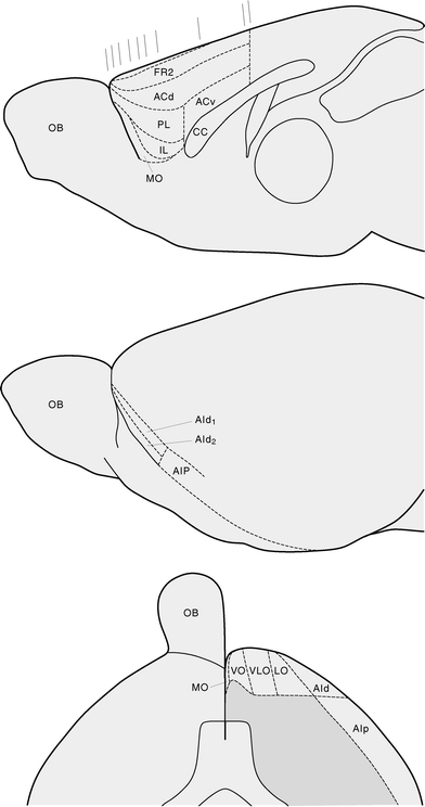
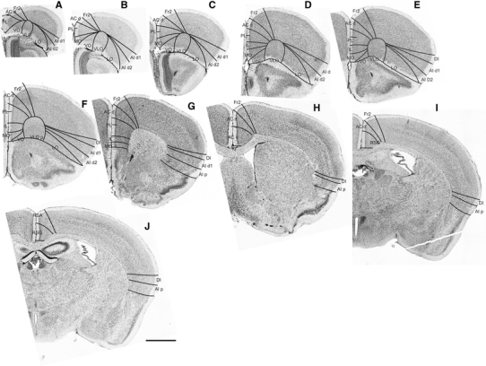
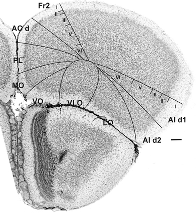
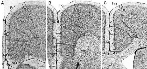
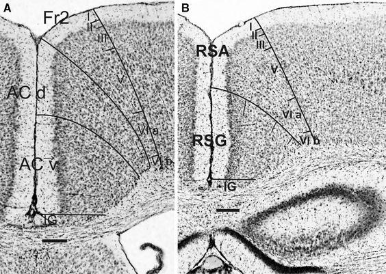
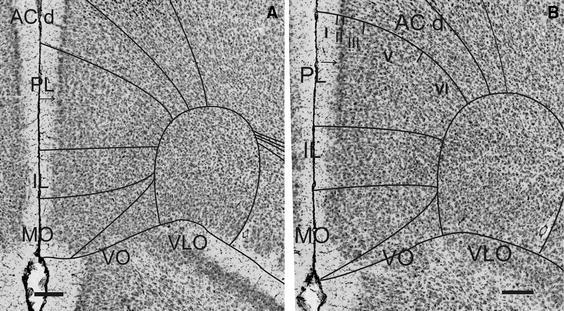
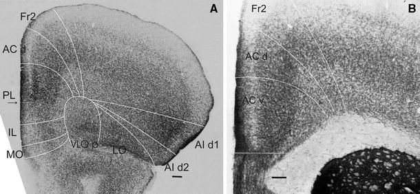
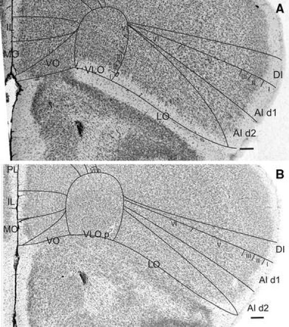
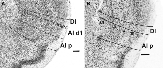
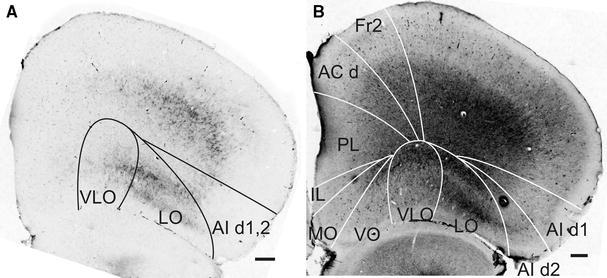
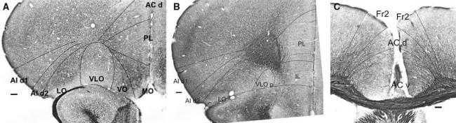
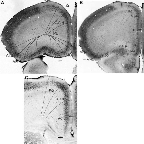
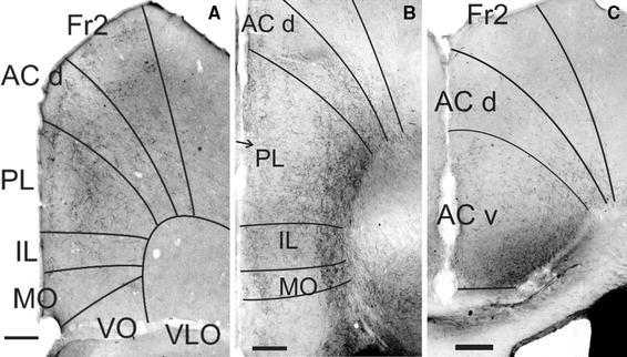
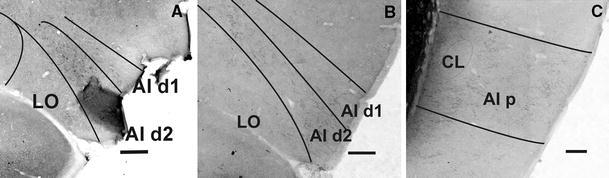
This study describes cytoarchitectonic criteria to define the prefrontal cortical areas in the mouse brain (C57BL/6 strain). Currently, well-illustrated mouse brain stereotaxic atlases are available, which, however, do not provide a description of the distinctive cytoarchitectonic characteristics of individual prefrontal areas. Such a description is of importance for stereological, neuronal tracing, and physiological, molecular and neuroimaging studies in which a precise parcellation of the prefrontal cortex (PFC) is required. The present study describes and illustrates: the medial prefrontal areas, i.e., the infralimbic, prelimbic, dorsal and ventral anterior cingulate and Fr2 area; areas of the lateral PFC, i.e., the dorsal agranular insular cortical areas and areas of the ventral PFC, i.e., the lateral, ventrolateral, ventral and medial orbital areas. Each cytoarchitectonically defined boundary is corroborated by one or more chemoarchitectonic stainings, i.e., acetylcholine esterase, SMI32, SMI311, dopamine, parvalbumin, calbindin and myelin staining.
Figures

References
-
- Cavada C, Comañy T, Hernández-González A, Reinoso-Suárez F. Acetylcholinesterase histochemistry in the macaque thalamus reveals territories selectively connected to frontal, parietal and temporal association cortices. J Chem Neuroanat. 1995;8:245–257. doi: 10.1016/0891-0618(95)00050-H. - DOI - PubMed
Publication types
MeSH terms
Substances
Grants and funding
LinkOut - more resources
Full Text Sources
Miscellaneous
