. 2010 May;19(5):987-99.
doi: 10.1002/pro.376.

Structure of the catalytic domain of the human mitochondrial Lon protease: proposed relation of oligomer formation and activity

Affiliations

Structure of the catalytic domain of the human mitochondrial Lon protease: proposed relation of oligomer formation and activity

Javier García-Nafría et al. Protein Sci. 2010 May.

Abstract

ATP-dependent proteases are crucial for cellular homeostasis. By degrading short-lived regulatory proteins, they play an important role in the control of many cellular pathways and, through the degradation of abnormally misfolded proteins, protect the cell from a buildup of aggregates. Disruption or disregulation of mammalian mitochondrial Lon protease leads to severe changes in the cell, linked with carcinogenesis, apoptosis, and necrosis. Here we present the structure of the proteolytic domain of human mitochondrial Lon at 2 A resolution. The fold resembles those of the three previously determined Lon proteolytic domains from Escherichia coli, Methanococcus jannaschii, and Archaeoglobus fulgidus. There are six protomers in the asymmetric unit, four arranged as two dimers. The intersubunit interactions within the two dimers are similar to those between adjacent subunits of the hexameric ring of E. coli Lon, suggesting that the human Lon proteolytic domain also forms hexamers. The active site contains a 3(10) helix attached to the N-terminal end of alpha-helix 2, which leads to the insertion of Asp852 into the active site, as seen in M. jannaschii. Structural considerations make it likely that this conformation is proteolytically inactive. When comparing the intersubunit interactions of human with those of E. coli Lon taken with biochemical data leads us to propose a mechanism relating the formation of Lon oligomers with a conformational shift in the active site region coupled to a movement of a loop in the oligomer interface, converting the proteolytically inactive form seen here to the active one in the E. coli hexamer.


Figures
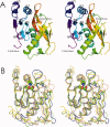
Figure 1
Figure 1
The hLonP protomer. A: Stereo ribbon diagram color ramped from N- to C-terminus. B: In the same orientation as (A), the structures of E. coli (coral), M. janaschii (blue), and A. fulgidus (yellow) LonP represented as worms superimposed on that of hLonP (green), confirming their high similarity. The main differences are in the loops and at the N-terminus of α2. Ser855 and Lys898 composing the catalytic dyad are shown as spheres for the human enzyme. This Figure and Figures 3 and 4 were created with CCP4mg.
Figure 2
Figure 2
Sequence alignment of the Lon proteolytic domains for which the structure is known. The secondary structure at the top corresponds to the human orthologue, whereas that at the bottom corresponds to EcLonP. Invariant positions are shaded and conserved positions are boxed. Figure created using CLUSTAL W and ESPript.
Figure 3
Figure 3
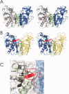
The subunit–subunit interface in LonP and relation between oligomerization and activation. A: Stereo view of the hLonP dimer interface, formed by chains (A) (gray) and (B) (blue) shown as ribbons. α1 of one chain packs against β-sheet 1 of the adjacent subunit. Side chains (cylinders with carbons in green) are shown for those residues involved in direct intersubunit contacts together with the H-bonds between them. The loop emerging from β4 (black) does not make any contacts with the adjacent subunit. Residues in the active site are depicted as cyan cylinders. B: Stereo view of adjacent subunits from the E. coli LonP hexamer colored coral and yellow. The side chains are shown as cylinders (green) for those residues involved in direct intersubunit contacts. The extended loop (residues 669–674)—caused by the unfolding of the beginning of α2 in hLon—is in red, and makes additional contacts with the adjacent subunit. Residues in the active site are depicted as cyan cylinders. C: Structural differences between the monomeric/dimeric inactive state (represented by hLon) and the proposed hexameric active state (represented by EcLon). The surfaces of two adjacent subunits of the EcLonP hexamer are shown in blue and white: the extensive surface buried in this interface is evident. For EcLonP, the following features are shown. (i) The extended loop residues 667–681 (including a length of β-strand) at the N-terminal end of α2 (red ribbon). This makes a substantial contribution to the interface. (ii) The side chains (red cylinders) of two key residues Asp676 and Trp603 (equivalent to Asp852 and Trp770 in hLonP). For hLonP are shown: (i) the catalytic residues Ser855, Lys898, and Thr880 (cylinders colored by atom type), (ii) the N-terminal segment of hLonP α2 as a black ribbon, (iii) the side chains of the two residues as black cylinders (Asp852 and Trp770) which block the active site, and (iv) the chain in the neighborhood of the catalytic serine as a pale green ribbon. The hLonP N-terminal segment of α2 with the helical fold places Asp852 directly in the active site with Trp770 physically blocking access, making the monomeric/dimeric form inactive. Thus, in EcLonP, the N-terminal section of α2 (which is helical in hLon) is unwound so as to contact the adjacent subunit. This movement carries Asp676 (equivalent to Asp852 in human) away from the active site. The loop is H-bonded to the main chain of Trp603 whose side chain is also pulled away from the active site.
Figure 4
Figure 4
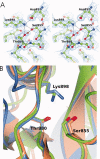
A: Active site of the hLon proteolytic domain with the 2FoFc electron density contoured at 1σ. The hydrogen bond network between the catalytic nucleophile Ser855, the general base Lys898 and Thr880 is shown. Asp852 can be seen to prevent access of substrate by H-bonding to Lys898. B: Superposition of the region around the active site of the hLonP domain (green) with those of the VP4 proteases from the infectious pancreatic necrosis virus (2pnl, coral) and the blotched snakehead virus (2gef, blue). The positions of the two residues of the catalytic dyad and the associated threonine are closely similar in the three structures.

Similar articles

Cited by

References

    1. Gottesman S, Maurizi MR. Regulation by proteolysis: energy-dependent proteases and their targets. Microbiol Rev. 1992;56:592–621. - PMC - PubMed
    1. Schoemaker JM, Markovitz A. Identification of the gene Lon (capR) product as a 94-kilodalton polypeptide by cloning and deletion analysis. J Bacteriol. 1981;147:46–56. - PMC - PubMed
    1. Ostersetzer O, Kato Y, Adam Z, Sakamoto W. Multiple intracellular locations of Lon protease in Arabidopsis: evidence for the localization of AtLon4 to chloroplasts. Plant Cell Physiol. 2007;48:881–885. - PubMed
    1. Wang N, Gottesman S, Willingham MC, Gottesman MM, Maurizi MR. A human mitochondrial ATP-dependent protease that is highly homologous to bacterial Lon protease. Proc Natl Acad Sci USA. 1993;90:11247–11251. - PMC - PubMed
    1. Kikuchi M, Hatano N, Yokota S, Shimozawa N, Imanaka T, Taniguchi H. Proteomic analysis of rat liver peroxisome: presence of peroxisome-specific isozyme of Lon protease. J Biol Chem. 2004;279:421–428. - PubMed

Publication types

LinkOut - more resources