Review
. 2010 Jun;90(6):655-63.
doi: 10.1016/j.exer.2010.02.014. Epub 2010 Mar 16.

Membrane-tethered mucins have multiple functions on the ocular surface

Affiliations
Review

Membrane-tethered mucins have multiple functions on the ocular surface

Bharathi Govindarajan et al. Exp Eye Res. 2010 Jun.

Abstract

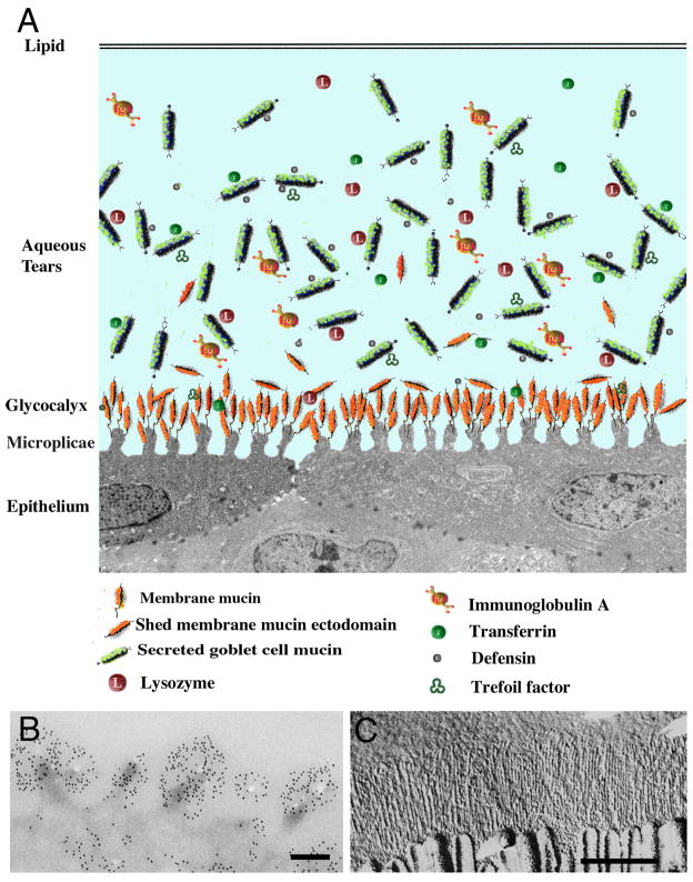
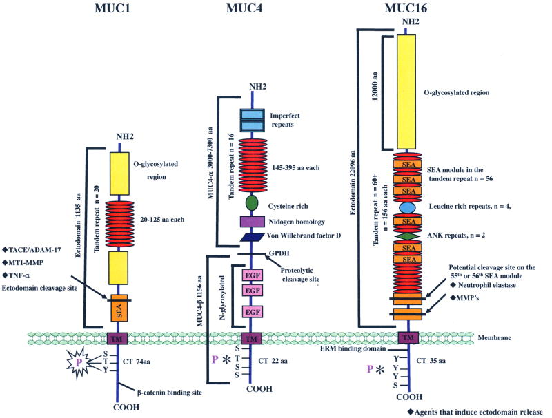
Membrane-tethered mucins are large glycoproteins present in the glycocalyx along the apical surface of all wet-surfaced epithelia of the body, including that of the ocular surface. Originally thought to function only in epithelial surface lubrication and hydration, data now indicate that the mucins are multifunctional molecules, each having unique as well as common functions. This review summarizes current knowledge regarding the three major membrane mucins of the ocular surface, MUC1, MUC4, and MUC16. The mucins vary in their ocular surface distribution, size, structural motifs, and functions. The ectodomains of each are released into the tear film and are, thus, a component of the soluble mucins of the tear film. Both animal and in vitro models for their study are herein described, as are alterations of the mucins in ocular surface disease.


Figures

Fig. 1
Fig. 1
Diagram and electron micrograph images of the tear film-glycocalyx interface. (A) Diagram of the tear film demonstrating tear components and the apical surface glycocalyx with its membrane-tethered mucins (after (Gipson, I.K., 2004)). (B, C) Transmission electron micrograph showing immunogold localization of MUC16 on microplicae. Bar = 0.25 μm. (C) Freeze-fracture image demonstrating long filamentous structure within the glycocalyx of the gut epithelium that appear, based on length, to be membrane-tethered mucins (from (Swift, J.G. and Mukherjee, T.M., 1976)). Bar = 0.5 μm.
Fig. 2
Fig. 2
Diagram of the molecular domains of the major ocular surface, membrane-tethered mucins MUC1, MUC4 and MUC16. The membrane mucins represented share a heavily O-glycosylated ectodomain, comprised of a variable number of tandem repeats rich in serine, threonine, and proline. The amino terminal domains of MUC1 and MUC16 have a non tandem repeat, whereas the tandem repeat domain of MUC4 extends to the NH2 terminus. MUCs 1 and 16 have 1 and 56 SEA modules, respectively, while MUC4 has none. MUC4 is the only membrane-tethered mucin that contains EGF domains. The ectodomain release sites of each mucin are indicated by an arrow, and agents that have been demonstrated to induce ectodomain release are indicated by ◆. The cytoplasmic tails (CT) of the three membrane mucins differ in length and binding partners. The cytoplasmic tail of MUC1 can be phosphorylated and contains a β-catenin binding site. The potential phosphorylation sites on MUCs 4 and 16 are indicated by *. MUC16 CT, along with potential phosphorylation, sites has an ezrin/radixin/moesin binding domain.
Fig. 3
Fig. 3
Rose bengal dye penetrance in cultured stratified HCLE cells with or without knockdown of MUC16 using siRNA methods. The untreated control and vector only control show island of cells that exclude rose bengal dye, whereas cells with MUC16 knockdown, either siRNA sequence 1 or 2, do not exclude the dye. These data demonstrate the role of the membrane-tethered mucin MUC16 in barrier function. Original data in Blalock et al., 2007 (Blalock, T.D. et al., 2007). Bar = 50 um.

Similar articles

Cited by

References

    1. Albertsmeyer A, Kakkassery V, Spurr-Michaud S, Beeks O, Gipson I. Effect of pro-inflammatory mediators on membrane-associated mucins expressed by human ocular surface epithelial cells. Exp Eye Res In press. - PMC - PubMed
    1. Araki-Sasaki K, Ohashi Y, Sasabe T, Hayashi K, Watanabe H, Tano Y, Handa H. An SV40-immortalized human corneal epithelial cell line and its characterization. Invest Ophthalmol Vis Sci. 1995;36:614–621. - PubMed
    1. Arango ME, Li P, Komatsu M, Montes C, Carraway CA, Carraway KL. Production and localization of Muc4/sialomucin complex and its receptor tyrosine kinase ErbB2 in the rat lacrimal gland. Invest Ophthalmol Vis Sci. 2001;42:2749–2756. - PubMed
    1. Argueso P, Balaram M, Spurr-Michaud S, Keutmann HT, Dana MR, Gipson IK. Decreased levels of the goblet cell mucin MUC5AC in tears of patients with Sjogren syndrome. Invest Ophthalmol Vis Sci. 2002;43:1004–1011. - PubMed
    1. Argueso P, Guzman-Aranguez A, Mantelli F, Cao Z, Ricciuto J, Panjwani N. Association of cell surface mucins with galectin-3 contributes to the ocular surface epithelial barrier. J Biol Chem. 2009;284:23037–23045. - PMC - PubMed