. 2010 Jun 2;167(4):1205-15.
doi: 10.1016/j.neuroscience.2010.03.002. Epub 2010 Mar 9.

Tooth pulp inflammation increases brain-derived neurotrophic factor expression in rodent trigeminal ganglion neurons

Affiliations

Tooth pulp inflammation increases brain-derived neurotrophic factor expression in rodent trigeminal ganglion neurons

L Tarsa et al. Neuroscience. .

Abstract

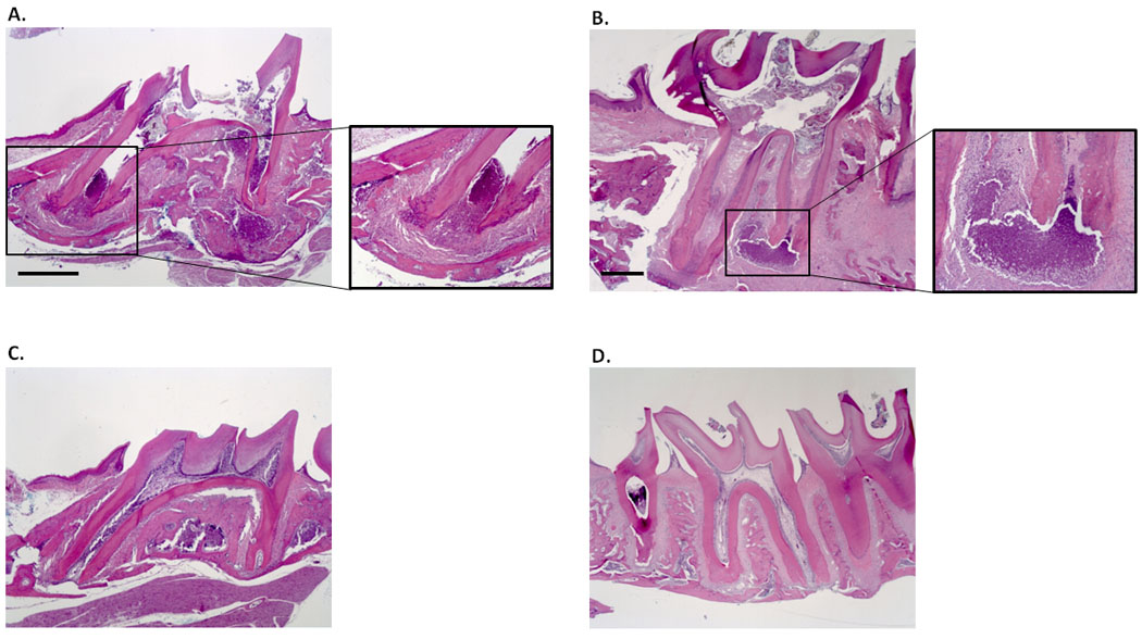
Nociceptive pathways with first-order neurons located in the trigeminal ganglion (TG) provide sensory innervation to the head, and are responsible for a number of common chronic pain conditions, including migraines, temporomandibular disorders and trigeminal neuralgias. Many of those conditions are associated with inflammation. Yet, the mechanisms of chronic inflammatory pain remain poorly understood. Our previous studies show that the neurotrophin brain-derived neurotrophic factor (BDNF) is expressed by adult rat TG neurons, and released from cultured newborn rat TG neurons by electrical stimulation and calcitonin gene-related peptide (CGRP), a well-established mediator of trigeminal inflammatory pain. These data suggest that BDNF plays a role in activity-dependent plasticity at first-order trigeminal synapses, including functional changes that take place in trigeminal nociceptive pathways during chronic inflammation. The present study was designed to determine the effects of peripheral inflammation, using tooth pulp inflammation as a model, on regulation of BDNF expression in TG neurons of juvenile rats and mice. Cavities were prepared in right-side maxillary first and second molars of 4-week-old animals, and left open to oral microflora. BDNF expression in right TG was compared with contralateral TG of the same animal, and with right TG of sham-operated controls, 7 and 28 days after cavity preparation. Our ELISA data indicate that exposing the tooth pulp for 28 days, with confirmed inflammation, leads to a significant upregulation of BDNF in the TG ipsilateral to the affected teeth. Double-immunohistochemistry with antibodies against BDNF combined with one of nociceptor markers, CGRP or transient receptor potential vanilloid type 1 (TRPV1), revealed that BDNF is significantly upregulated in TRPV1-immunoreactive (IR) neurons in both rats and mice, and CGRP-IR neurons in mice, but not rats. Overall, the inflammation-induced upregulation of BDNF is stronger in mice compared to rats. Thus, mouse TG provides a suitable model to study molecular mechanisms of inflammation-dependent regulation of BDNF expression in vivo.


Figures

Figure 1
Figure 1
Photomicrographs of hematoxylin/eosin-stained sagittal sections through the first right (A–C) and left (D) maxillary molar of a mouse (A, C) and rat (B, D). A cavity was prepared on the right side at 4 weeks of age, and was left open to oral microflora for 7 days (A) and 28 days (B). The periapical region shows infiltrates of neutrophils and pulpal abscesses (A, B; insets), which are absent in a sham operated animal (C) and on the intact left side (D). Scale bars, 500 µm.
Figure 2
Figure 2
Levels of BDNF protein (per gram of tissue) in left (Control) and right (Inflammation) trigeminal ganglia, seven and 28 days after the open cavity preparation in first and second maxillary molars on the right side in (A) rats (7 days, n=8 rats; 28 days, n=11 rats), and (B) mice (7 days, n=3 mice; 28 days, n=4 mice); * P<0.05, ** P<0.01.
Figure 3
Figure 3
(A) Micrographs of a single 10-µm section of the trigeminal ganglion from a 5-week-old rat double-immunostained for BDNF (αBDNF) and TRPV1 (αTRPV1). An overlay image (Overlay) shows that many TRPV1-immunoreactive cells also express BDNF (arrows). Scale bar, 100 µm. (B) Mean density of BDNF/TRPV1 double-immunoreactive (IR) cells in left (Control) and right (Inflammation) trigeminal ganglia from 5-week-old rats (left panel) and mice (right panel) with cavities prepared in right-side first and second molars at 4-weeks of age. (C) Mean density of all (i.e., BDNF-IR and BDNF-non-IR) TRPV1-IR cells counted in the same microscope fields as BDNF/TRPV1-IR cells presented in panel B. Numbers of analyzed cells: Rat, 7 days: Control, 1789 TRPV1-IR and 757 BDNF/TRPV1-IR, Inflammation, 2000 TRPV1-IR and 998 BDNF/TRPV1-IR (N=2 rats); 28 days: Control, 1211 TRPV1-IR and 591 BDNF/TRPV1-IR, Inflammation, 2332 TRPV1-IR and 1226 BDNF/TRPV1-IR (N=3 rats). Mouse, 7 days: Control, 438 TRPV1-IR and 139 BDNF/TRPV1-IR, Inflammation, 1967 TRPV1-IR and 758 BDNF/TRPV1-IR; 28 days: Control, 4792 TRPV1-IR and 1160 BDNF/TRPV1-IR, Inflammation, 6508 TRPV1-IR and 2644 BDNF/TRPV1-IR. (N=3 mice); * P<0.05; *** P<0.001.
Figure 4
Figure 4
(A) Micrographs of a single 10-µm section of the trigeminal ganglion from a 5-week-old mouse double-immunostained for BDNF (αBDNF) and CGRP (αCGRP). An overlay image (Overlay) shows that some CGRP-positive cells also show BDNF immunoreactivity (arrows). Scale bar, 50 µm. (B) Mean density of BDNF/CGRP double-immunoreactive (IR) cells in left (Control) and right (Inflammation) trigeminal ganglia from 5-week-old rats (left panel) and mice (right panel) with cavities prepared in right-side first and second molars at 4-weeks of age. (C) Mean density of all (i.e., BDNF-IR and BDNF-non-IR) CGRP-IR cells counted in the same fields as BDNF/CGRP-IR cells presented in panel B. The analysis of BDNF/CGRP-IR cells was performed on alternate sections of the ganglia used for the analysis of BDNF/TRPV1-IR cells presented in Figure 3. Numbers of analyzed cells: Rat, 7 days: Control, 2450 CGRP-IR and 440 BDNF/CGRP-IR, Inflammation, 2550 CGRP-IR and 474 BDNF/CGRP-IR (N=2 rats); 28 days: Control, 2642 CGRP-IR and 628 BDNF/CGRP-IR, Inflammation, 3344 CGRP-IR and 654 BDNF/CGRP-IR (N=3 rats). Mouse, 7 days: Control, 1490 CGRP-IR and 548 BDNF/CGRP-IR, Inflammation, 1262 CGRP-IR and 442 BDNF/CGRP-IR; 28 days: Control, 3079 CGRP-IR and 699 BDNF/CGRP-IR, Inflammation, 2967 CGRP-IR and 838 BDNF/CGRP-IR. (N=3 mice); *** P<0.001; ns, non-significant.

Similar articles

Cited by

References

    1. Aid T, Kazantseva A, Piirsoo M, Palm K, Timmusk T. Mouse and rat BDNF gene structure and expression revisited. J Neurosci Res. 2007;85:525–535. - PMC - PubMed
    1. Amaya F, Shimosato G, Nagano M, Ueda M, Hashimoto S, Tanaka Y, Suzuki H, Tanaka M. NGF and GDNF differentially regulate TRPV1 expression that contributes to development of inflammatory thermal hyperalgesia. Eur J Neurosci. 2004;20:2303–2310. - PubMed
    1. Balkowiec A, Bałkowiec-Iskra E. Novel Approaches to Studying Activity-Dependent Regulation of Neurotrophins and Neuropeptides in Sensory Pathways from Orofacial Tissues. In: Daskalaki A, editor. Informatics in Oral Medicine: Advanced Techniques in Clinical and Diagnostic Technologies. IGI Global; 2010.
    1. Balkowiec A, Kunze DL, Katz DM. Brain-derived neurotrophic factor acutely inhibits AMPA-mediated currents in developing sensory relay neurons. J Neurosci. 2000;20:1904–1911. - PMC - PubMed
    1. Bałkowiec-Iskra E, Balkowiec A. Regulation of BDNF expression in trigeminal ganglion neurons by proinflammatory cytokines. Soc Neurosci Abstr. 2009 710.4.

Publication types

Substances