. 2010 Apr;137(8):1231-41.
doi: 10.1242/dev.042051. Epub 2010 Mar 10.

The Mi-2-like Smed-CHD4 gene is required for stem cell differentiation in the planarian Schmidtea mediterranea

Affiliations

The Mi-2-like Smed-CHD4 gene is required for stem cell differentiation in the planarian Schmidtea mediterranea

M Lucila Scimone et al. Development. 2010 Apr.

Abstract

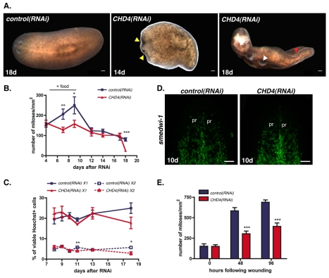
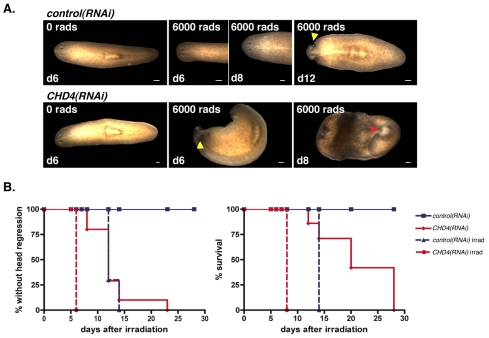
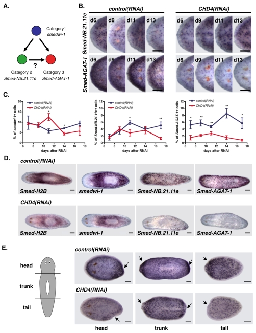
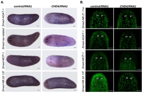
Freshwater planarians are able to regenerate any missing part of their body and have extensive tissue turnover because of the action of dividing cells called neoblasts. Neoblasts provide an excellent system for in vivo study of adult stem cell biology. We identified the Smed-CHD4 gene, which is predicted to encode a chromatin-remodeling protein similar to CHD4/Mi-2 proteins, as required for planarian regeneration and tissue homeostasis. Following inhibition of Smed-CHD4 with RNA interference (RNAi), neoblast numbers were initially normal, despite an inability of the animals to regenerate. However, the proliferative response of neoblasts to amputation or growth stimulation in Smed-CHD4(RNAi) animals was diminished. Smed-CHD4(RNAi) animals displayed a dramatic reduction in the numbers of certain neoblast progeny cells. Smed-CHD4 was required for the formation of these neoblast progeny cells. Together, these results indicate that Smed-CHD4 is required for neoblasts to produce progeny cells committed to differentiation in order to control tissue turnover and regeneration and suggest a crucial role for CHD4 proteins in stem cell differentiation.


Figures

Fig. 1.
Fig. 1.
CHD4 is required for planarian regeneration and is expressed in neoblasts. (A) As shown in comparison with a control animal, a CHD4(RNAi) worm did not regenerate 7 days following amputation (41/41 were similar). Worms were fed three times and amputated at day 8 after initial RNAi. White dotted line represents the blastema boundary. Anterior is to the left. (B) Expression of CHD4 in wild-type and irradiated (6000 rads 5 days prior) animals, dorsal and ventral views (7/7 were similar). Expression of an irradiation-insensitive gene, PC2 (expressed in the nervous system) served as a control. Anterior is to the left. (C) Expression of CHD4 in neoblast subsets, X1 (1472 cells counted) and X2 (655 cells counted), and radiation-insensitive cells, Xins (1117 cells counted). Scale bars: 0.1 mm.
Fig. 2.
Fig. 2.
CHD4 is required for homeostasis and normal stimulation of neoblast proliferation. (A) Control and CHD4(RNAi) animals during normal tissue turnover 14 and 18 days after initial RNAi. Yellow arrowhead, head regression; white arrowhead, lesions; red arrowhead, curling (10/10 were similar). Anterior is to the left. (B) Numbers of mitoses labeled with an anti-phospho histone 3 (αH3P) antibody were divided by animal surface area. Animals were fed three times and fixed at time points after initial RNAi. ‘+ food’ indicates the proliferation response following feedings. Data are means ± s.e.m. (n=2 experiments, >8 worms per time point). *, P<0.05; **, P<0.001, ***, P<0.0001, Student’s t-test. (C) Animals were fed three times with dsRNA and cell macerates were generated at multiple time points after initial RNAi. Cells were labeled with Hoechst 33342, calcein, and propidium iodide; X1 and X2 cell percentages (in the total population of Hoechst-labeled cells) were determined by flow cytometry. Data are means ± s.e.m. (n=3 experiments). *, P<0.05; **, P<0.001, Student’s t-test. (D) Fluorescence in situ hybridizations of animals fixed 10 days after multiple dsRNA feedings using the smedwi-1 riboprobe (>12 worms per condition were similar). pr, photoreceptors. Anterior is up. (E) Numbers of mitoses labeled with αH3P were divided by animal surface area. Animals were fed three times, amputated 8 days after initial feeding and fixed at different times following wounding. Data are means ± s.e.m.; n=2 experiments, >8 animals per time point. ***, P<0.0001, Student’s t-test. Scale bars: 0.1 mm.
Fig. 3.
Fig. 3.
CHD4(RNAi) intact animals die faster following irradiation. (A) Control and CHD4(RNAi) animals were lethally irradiated with 6000 rads 8 days following initial RNAi. Irradiated control(RNAi) animals showed head regression by 12 days following irradiation (7/10 animals) and curling by 14 days (10/10), whereas irradiated CHD4(RNAi) animals showed head regression by 6 days following irradiation (10/10) and curling by 8 days (10/10). Yellow arrowhead, head regression; red arrowhead, curling. Anterior is to the left. (B) Percentages of control or CHD4(RNAi) animals without head regression (left) and still alive (right) following irradiation are shown. Scale bars: 0.1 mm.
Fig. 4.
Fig. 4.
CHD4(RNAi) animals have reduced numbers of Smed-AGAT-1-expressing cells. (A) Lineage-related cells and the markers expressed in each cell type. Other models are possible, see Eisenhoffer et al. (Eisenhoffer et al., 2008). (B) Head regions of whole-mount in situ hybridizations using a Category 2 probe Smed-NB.21.11e (upper row) and a Category 3 probe Smed-AGAT-1 (lower row) of control and CHD4(RNAi) animals at several time points following initial RNAi (>4 worms/time point displayed similar results). Anterior is to the left. (C) Cell in situ hybridizations using a Category 1 probe (smedwi-1; >823 cells per time point), a Category 2 probe (Smed-NB.21.11e; >954 cells per time point) and a Category 3 probe (Smed-AGAT-1; >1157 cells per time point) of control and CHD4(RNAi) worm macerates at multiple times after initial RNAi. Percentages of smedwi-1+, Smed-NB.21.11e+, and Smed-AGAT-1+ cells within the total DAPI-positive cell population are shown (means ± s.e.m., n=3 experiments). *, P<0.05; **, P<0.001, Student’s t-test. (D) Whole-mount in situ hybridizations with Category 1 probes (Smed-H2B and smedwi-1), a Category 2 probe (Smed-NB.21.11e) and a Category 3 probe (Smed-AGAT-1) of control and CHD4(RNAi) animals 20 days following initial RNAi. At this late time point, CHD4(RNAi) animals displayed head regression, had lesions and began to curl (>7 worms displayed similar results). Anterior is to the left. (E) Whole-mount in situ hybridizations with the Smed-AGAT-1 riboprobe of control and CHD4(RNAi) head, trunk and tail pieces fixed 3 days following amputation. Animals were amputated 7 days following initial RNAi (>11 regenerating pieces displayed similar results). Arrows indicate the site of amputation. Anterior is to the left. Scale bars: 0.1 mm.
Fig. 5.
Fig. 5.
CHD4 is required for the formation of Smed-AGAT-1-expressing cells. (A) Smed-AGAT-1+ cell numbers normalized by animal length from whole-mount in situ hybridizations are shown at time points following irradiation. Animals (>4 worms per group) were fed once and lethally irradiated the same day (left) or fed three times and lethally irradiated at day 7 (right). Data are means ± s.e.m. (B) Percentage of double-labeled SMEDWI-1; Smed-AGAT-1-expressing cells in the total number of Smed-AGAT-1+ cells in control and CHD4(RNAi) animals (upper graph), and the percentage of double-labeled SMEDWI-1; Smed-AGAT-1-expressing cells normalized by the area of the head region (lower graph). Data are means ± s.e.m. (>5 worms per group). *, P<0.05; **, P<0.001; ***, P<0.0001, Student’s t-test. Animals were fed twice and fixed at time points following the initial RNAi. Fluorescence in situ hybridizations using Smed-AGAT-1 riboprobe (green) and SMEDWI-1 antibody (red) are shown. Arrowheads indicate cells that co-express Smed-AGAT-1 and SMEDWI-1. (C) SMEDWI-1-expressing cell numbers in control and CHD4(RNAi) animals. pr, photoreceptors. Circled area, region counted. Data are means ± s.e.m. (>8 worms per group). **, P<0.001, Student’s t-test. A second experiment had similar results. Animals were fed twice and fixed at time points following initial RNAi. Anterior is up. Scale bars: 0.1 mm.
Fig. 6.
Fig. 6.
CHD4(RNAi) animals have decreased numbers of several Category 3-expressing cells. (A) Whole-mount in situ hybridizations using Category 3 probes 10 days following initial RNAi (>4 worms per riboprobe were similar). Anterior is to the left. (B) Fluorescence in situ hybridizations of animals fixed 10 days after multiple dsRNA feedings using the Smed-NB.21.11e, Smed-AGAT-1, Smed-MCP-1 and Smed-NB.52.12f riboprobes (>12 worms per condition were similar). The head region is shown. pr, photoreceptors. Anterior is up. Scale bars: 0.1 mm.

Similar articles

Cited by

References

    1. Ahringer J. (2000). NuRD and SIN3 histone deacetylase complexes in development. Trends Genet. 16, 351-356 - PubMed
    1. Baguñà J. (1976). Mitosis in the intact and regenerating planarian Dugesia mediterranea n.sp. I. Mitotic studies during growth, feeding and starvation. J. Exp. Zool. 195, 53-64
    1. Bardeen C. R., Baetjer F. H. (1904). The inhibitive action of the Roentgen rays on regeneration in planarians. J. Exp. Zool. 1, 191-195
    1. Becker P. B., Horz W. (2002). ATP-dependent nucleosome remodeling. Annu. Rev. Biochem. 71, 247-273 - PubMed
    1. Bibikova M., Laurent L. C., Ren B., Loring J. F., Fan J. B. (2008). Unraveling epigenetic regulation in embryonic stem cells. Cell Stem Cell 2, 123-134 - PubMed

Publication types