. 2010 Jul;38(13):4337-48.
doi: 10.1093/nar/gkq136. Epub 2010 Mar 11.

Genetic instability triggered by G-quadruplex interacting Phen-DC compounds in Saccharomyces cerevisiae

Affiliations

Genetic instability triggered by G-quadruplex interacting Phen-DC compounds in Saccharomyces cerevisiae

Aurèle Piazza et al. Nucleic Acids Res. 2010 Jul.

Abstract

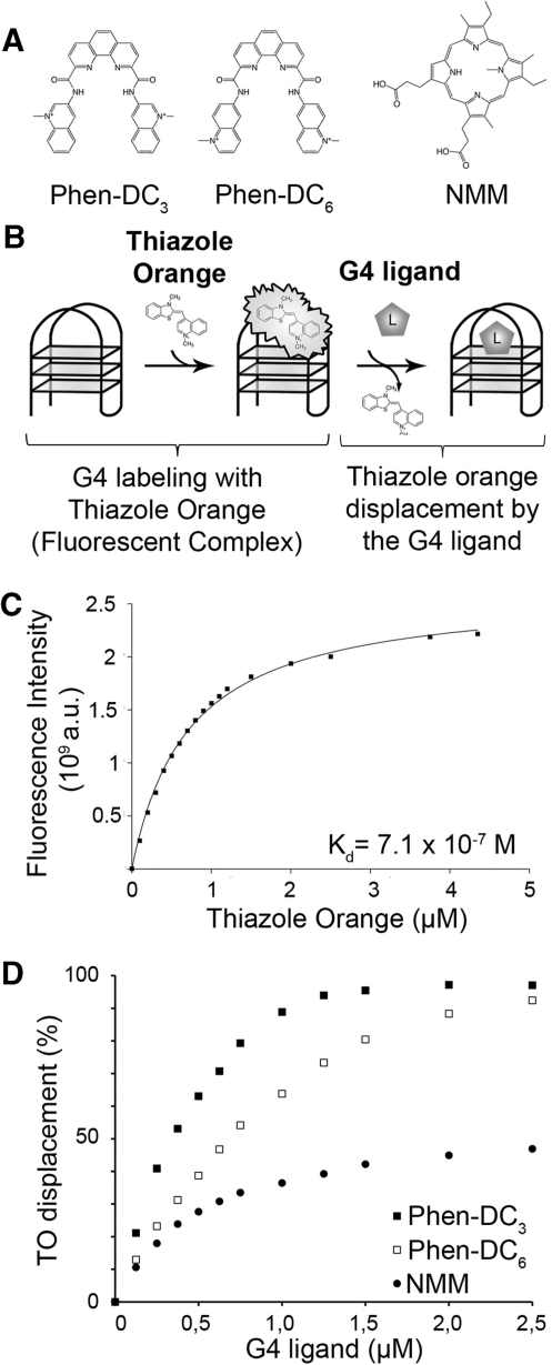
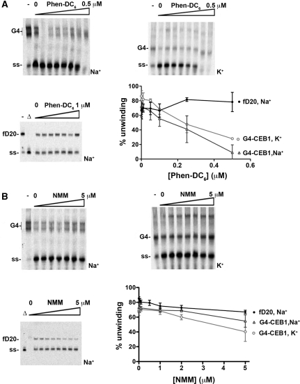
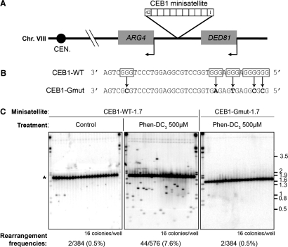
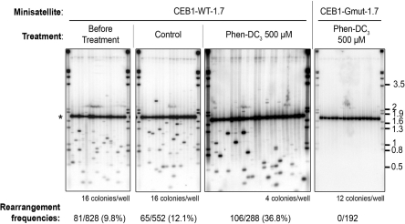
G-quadruplexes are nucleic acid secondary structures for which many biological roles have been proposed but whose existence in vivo has remained elusive. To assess their formation, highly specific G-quadruplex ligands are needed. Here, we tested Phen-DC(3) and Phen-DC(6), two recently released ligands of the bisquinolinium class. In vitro, both compounds exhibit high affinity for the G4 formed by the human minisatellite CEB1 and inhibit efficiently their unwinding by the yeast Pif1 helicase. In vivo, both compounds rapidly induced recombination-dependent rearrangements of CEB1 inserted in the Saccharomyces cerevisiae genome, but did not affect the stability of other tandem repeats lacking G-quadruplex forming sequences. The rearrangements yielded simple-deletion, double-deletion or complex reshuffling of the polymorphic motif units, mimicking the phenotype of the Pif1 inactivation. Treatment of Pif1-deficient cells with the Phen-DC compounds further increased CEB1 instability, revealing additional G4 formation per cell. In sharp contrast, the commonly used N-methyl-mesoporphyrin IX G-quadruplex ligand did not affect CEB1 stability. Altogether, these results demonstrate that the Phen-DC bisquinolinium compounds are potent molecular tools for probing the formation of G-quadruplexes in vivo, interfere with their processing and elucidate their biological roles.


Figures

Figure 1.
Figure 1.
Fluorescent intercalator displacement of the G-quadruplex formed by the CEB1 motif (G4-CEB1) with Phen-DC3, Phen-DC6 and NMM. (A) Phen-DC3, Phen-DC6 and NMM structures. (B) Schematic representation of the FID assay. (C) TO association curve with G4-CEB1. (D) TO displacement by (filled square) Phen-DC3, (open square) Phen-DC6 or (filled circle) NMM in presence of K+.
Figure 2.
Figure 2.
Inhibition of G4-CEB1 unwinding by Phen-DC6 and NMM in vitro. (A) Pif1 unwinding of G4-CEB1 or forked DNA substrate (fD20) in presence of increasing concentrations of Phen-DC6. The G4-CEB1 substrates were formed in presence of Na+ or K+ as indicated. Quantifications (mean ± SD) from three independent experiments are shown. Open triangle indicates boiled substrate. Dash indicates absence of Pif1. Position of G4-CEB1 (G4) and unwound substrate (ss) are indicated. (B) Same as in (A), using NMM as a G4 ligand.
Figure 3.
Figure 3.
Phen-DC compounds trigger G-quadruplex-dependent CEB1 instability. (A) Schematic representation of the CEB1 minisatellite insertion in the chromosome VIII. (B) Sequences of CEB1-WT and CEB1-Gmut motifs. G-runs involved in G-quadruplex formation are boxed. Point mutations introduced in CEB1-Gmut motif and preventing G-quadruplex formation in vitro are shown in bold. (C) Southern blot analysis of WT cells carrying CEB1-WT-1.7 (strain AND1212-10D) or CEB1-Gmut-1.7 (AND1227-5C) after control (1% DMSO) or 500 µM Phen-DC3 treatment. The number of colonies analyzed and the rearrangement frequencies are indicated below each gel. Size markers (kb) are indicated on the right. The position of the parental minisatellite alleles (42 repeats, 1.7 kb) is indicated by an asterisk.
Figure 4.
Figure 4.
Treatment with Phen-DC3 increases the instability of CEB1-WT-1.7 in pif1Δ cells. Southern blot analysis of CEB1-WT-1.7 (AND1202-11A) or CEB1-Gmut-1.7 (AND1206-4C-D11P2) instability in pif1Δ cells, before and after control (1% DMSO) or 500 µM Phen-DC3 treatment. Other legends are as in Figure 3.
Figure 5.
Figure 5.
Sequence and size of CEB1 rearrangements produced upon treatment with Phen-DC3. (A) Schematic representation of the natural CEB1-1.8 allele and of 14 rearrangements produced upon treatment of WT cells (ORT2914) with 500 µM Phen-DC3. Nucleotide sequences are reported in Supplementary Figure S6. Chimerical motifs combining the polymorphisms of more than three parental motifs (called ‘Mix’ in Supplementary Figure S6) are shown as a white box. Sequences of the two red motifs in C7 have not been determined. (B) Schematic representation of 13 rearrangements produced upon treatment of pif1Δ cells (ORT4841) with 500 µM Phen-DC3. (C) Size distribution of CEB1-WT-1.7 and CEB1-1.8 rearrangements in WT (AND1212-10D and ORT2914), pif1Δ (AND1202-11A and ORT4841), pif1-K264A (ORT5087-5E) or rad27Δ (AND1218-1A and ORD6713-8D) cells, with or without treatment with 500 µM Phen-DC3. Datasets are from this study, except the size distribution of rearranged CEB1-WT-1.7 alleles in rad27Δ cells, obtained from ref. (8) (strain AND1218-1A). The n indicates the number of variants analyzed. The vertical gray lines indicate the size of the parental alleles. For each distribution, dark bar: median; gray bar: mean; box: distance between the first and third quantiles; whiskers: extreme values; white circles: outliers. P-values were obtained by comparing the samples with a Mann–Whitney–Wilcoxon statistical test.

Similar articles

Cited by

References

    1. Gellert M, Lipsett MN, Davies DR. Helix formation by guanylic acid. Proc. Natl Acad. Sci. USA. 1962;48:2013–2018. - PMC - PubMed
    1. Phan AT, Kuryavyi V, Luu KN, Patel DJ. Structural diversity of G-quadruplex scaffolds. In: Neidle S, Balasubramanian S, editors. Quadruplex Nucleic Acids. UK: Ch. 3, RSC publishing, Cambridge; 2006. pp. 81–99.
    1. Lipps HJ, Rhodes D. G-quadruplex structures: In vivo evidence and function. Trends Cell. Biol. 2009;19:414–422. - PubMed
    1. Maizels N. Dynamic roles for G4 DNA in the biology of eukaryotic cells. Nat. Struct. Mol. Biol. 2006;13:1055–1059. - PubMed
    1. Duquette ML, Handa P, Vincent JA, Taylor AF, Maizels N. Intracellular transcription of G-rich DNAs induces formation of G-loops, novel structures containing G4 DNA. Genes Dev. 2004;18:1618–1629. - PMC - PubMed

Publication types

MeSH terms