. 2010 May 14;285(20):15137-15148.
doi: 10.1074/jbc.M109.072843. Epub 2010 Mar 11.

Protease-activated receptor-1 (PAR1) acts via a novel Galpha13-dishevelled axis to stabilize beta-catenin levels

Affiliations

Protease-activated receptor-1 (PAR1) acts via a novel Galpha13-dishevelled axis to stabilize beta-catenin levels

Hagit Turm et al. J Biol Chem. .

Abstract

We have previously shown a novel link between hPar-1 (human protease-activated receptor-1) and beta-catenin stabilization. Although it is well recognized that Wnt signaling leads to beta-catenin accumulation, the role of PAR1 in the process is unknown. We provide here evidence that PAR1 induces beta-catenin stabilization independent of Wnt, Fz (Frizzled), and the co-receptor LRP5/6 (low density lipoprotein-related protein 5/6) and identify selective mediators of the PAR1-beta-catenin axis. Immunohistological analyses of hPar1-transgenic (TG) mouse mammary tissues show the expression of both Galpha(12) and Galpha(13) compared with age-matched control counterparts. However, only Galpha(13) was found to be actively involved in PAR1-induced beta-catenin stabilization. Indeed, a dominant negative form of Galpha(13) inhibited both PAR1-induced Matrigel invasion and Lef/Tcf (lymphoid enhancer factor/T cell factor) transcription activity. PAR1-Galpha(13) association is followed by the recruitment of DVL (Dishevelled), an upstream Wnt signaling protein via the DIX domain. Small interfering RNA-Dvl silencing leads to a reduction in PAR1-induced Matrigel invasion, inhibition of Lef/Tcf transcription activity, and decreased beta-catenin accumulation. It is of note that PAR1 also promotes the binding of beta-arrestin-2 to DVL, suggesting a role for beta-arrestin-2 in PAR1-induced DVL phosphorylation dynamics. Although infection of small interfering RNA-LRP5/6 or the use of the Wnt antagonists, SFRP2 (soluble Frizzled-related protein 2) or SFRP5 potently reduced Wnt3A-mediated beta-catenin accumulation, no effect was observed on PAR1-induced beta-catenin stabilization. Collectively, our data show that PAR1 mediates beta-catenin stabilization independent of Wnt. We propose here a novel cascade of PAR1-induced Galpha(13)-DVL axis in cancer and beta-catenin stabilization.


Figures

FIGURE 1.
FIGURE 1.
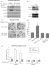
Activation of PAR1 induces nuclear β-catenin localization. ai, fluorescent immunostaining of β-catenin in HT-29 cells. Untreated control cells show weak staining of β-catenin (A). Cells treated with SFLLRN (100 μm) for 4 h show a somewhat stronger, yet still weak, level of staining using anti-β-catenin, detected with Cy3-conjugated goat anti-mouse secondary antibodies (B). A similar pattern is observed when the cells were pretreated for 2 h with 30 mm LiCl, but were non-activated for PAR1 (C). In contrast, a significant increase in nuclear β-catenin localization is seen in cells pretreated with LiCl and SFLLRN activated for 4 h. The arrows point to nuclear localization of β-catenin (D). E, phase-contrast image of the cells. aii, Western blot analysis of HT-29 cell nuclear fraction. The kinetics of HT-29 cell nuclear fraction shows a pattern of increased β-catenin levels following PAR1 activation. Maximal levels were obtained by 5 h after activation and remained high up to 20 h. b, PAR1 activation induces β-catenin stabilization in RKO cells. RKO cells were pretreated with LiCl (30 mm for 2 h) before (−) and after (4 and 5 h) PAR1 activation with thrombin. Whereas no β-catenin is detected prior to LiCl treatment, regardless of PAR1 activation, a marked increase in β-catenin level is seen following PAR1 activation after incubation with LiCl. c, immunohistochemical staining of hPar1-TG mammary gland tissues using both anti-Gα13 (a and b) and anti-Gα12 (c and d) antibodies. Mouse mammary glands were obtained from hPar1-TG mice targeted to overexpress in the mammary glands. Tissues from 12-day pregnancy show abundant localization of Gα13 and Gα12 compared with age-matched WT mouse mammary glands (magnification, ×20 (left) and ×40 (right)). The data are representative of four staining experiments.
FIGURE 2.
FIGURE 2.
13, but not Gα12, is involved in PAR1-mediated β-catenin stabilization. a, level of β-catenin. HEK-293 cells were transiently transfected with FLAG-β-catenin and either empty vector or constitutively active (Gα12QL and Gα13QL) plasmids. Following TFLLRNDK PAR1 activation, immunoblots were analyzed by using anti-FLAG (for FLAG-β-catenin) or anti-Gα12, anti-Gα13, and anti-β-actin antibodies. Gα13, but not Gα12, potently induced β-catenin stabilization. b, constitutively active Gα13-induced β-catenin stabilization is inhibited by a DN Gα13GA plasmid. HEK-293 cells were transiently transfected with FLAG-β-catenin and either constitutively active Gα13QL alone or with DN (Gα13GA) Gα13 plasmids. The DN Gα13GA form inhibited β-catenin stabilization induced by Gα13QL in a dose-dependent manner. ci, the Gα12 constructs, constitutively active (Gα12QL) and DN (Gα12GA), do not affect β-catenin stabilization. HEK-293 cells were transiently transfected with FLAG-β-catenin and combinations of either Gα12QL with Gα12GA or Gα12GA with Gα13QL as well as a combination of Gα13GA with Gα13QL. The Gα12 plasmid does not induce β-catenin stabilization, and there is no overlap between Gα12 and Gα13 plasmids in the induction of β-catenin stabilization. cii, representative histograms show the relative intensities of the bands expressed as a ratio between β-catenin and β-actin. The data are representative of three different experiments performed in triplicate. Error bars, ±S.D. *, p < 0.001. d, Gα12−/−/Gα13−/− MEFs fail to enhance β-catenin levels following PAR1 activation. Western blot analysis of both WT and Gα12−/−/Gα13−/− MEFs shows that thrombin activation (for 5 h) of the knock-out MEFs fails to induce β-catenin levels. In contrast, WT MEFs that express Gα13 (see middle panel) show enhanced β-catenin levels following PAR1 activation.
FIGURE 3.
FIGURE 3.
Effect of Rho and PLC-β inhibitors on HT-29 nuclear β-catenin levels. a, PAR1-induced nuclear accumulation of β-catenin is inhibited in the presence of Y27632, a Rho inhibitor (part of the Gα12/13 pathway), but not by U73122, a known inhibitor for PLC-β (a component of the Gαq pathway). The data are representative of three different experiments performed in triplicate. Error bars, ±S.D. *, p < 0.001. b, in contrast, upon Wnt3A stimulation, a potent reduction is seen in the presence of the inhibitor U3122 as compared with no effect obtained in the presence of the Y27632 inhibitor. Lower panel, quantification of the intensity of the bands per lamin as a control for nuclear protein loading was performed. The data are representative of three different experiment performed in triplicates. Error bars, ±S.D. *, p < 0.001.
FIGURE 4.
FIGURE 4.
The DN form of Gα13 inhibits both PAR1-induced Lef/Tcf transcriptional activity and Matrigel invasion. a, PAR1-induced Lef/Tcf transcriptional activity. TOPflash and FOPflash luciferase transcription activity was evaluated following PAR1 activation or in the presence of the constitutively active and DN Gα13 constructs. Although PAR1 activation (e.g. thrombin) elicits luciferase Lef/Tcf activity, the DN form inhibited it, and the constitutively active form markedly elevated it. The levels of TOPflash luciferase activity were compared with control FOPflash luciferase levels. S.D. was determined by Student's t test; *, p < 0.001; **, p < 0.005 (n = 3). b, the DN form of Gα13 inhibits Matrigel invasion. i, HEK-293 cells transfected with the Gα13GA construct display markedly reduced Matrigel invasion properties in response to PAR1 thrombin activation. ii, histograms show the number of invading cells. The data are representative of four independent experiments performed in triplicate. Error bars, ±S.D. The p value was determined. **, p < 0.005.
FIGURE 5.
FIGURE 5.
13 specifically interacts with DVL following PAR1 activation. a, activation of PAR1 induces Gα13-DVL interactions. HEK-293 cells were transiently transfected with FLAG-Dvl and WT Gα13. Immunoprecipitation analysis using anti-DVL antibodies was performed after PAR1 activation. Specific interaction between DVL and Gα13 is observed, reaching a maximum by 15 min of PAR1 activation. b, interaction between Gα13 and DVL is inhibited by PAR1-specific antagonist. HEK-293 cells were transiently transfected with FLAG-Dvl and WT Gα13. After PAR1 activation, SCH79797 immunoprecipitation by anti-Gα13 antibodies was performed in the presence or absence of a potent PAR1 antagonist. Specific association between DVL and Gα13 was observed. Maximal interaction was observed 15 min following PAR1 activation. Detection using anti-FLAG antibodies showed a potent inhibition of the association between DVL and Gα13 by the PAR1 antagonist. c, PAR1 activation induces DVL phosphorylation. The phosphorylation state of the DVL1 isoform was detected as a phosphorylation-dependent mobility shift of the DVL1 isoform. HEK-293 cells were transfected with FLAG-Dvl1 and either activated (e.g. thrombin) for the indicated periods of time or treated with Wnt3A (e.g. conditioned medium, 2 h). The levels of the phosphorylated form of DVL1 were increased after 0.5 h of PAR1 activation. di, cytosolic DVL1 is transferred to the plasma membrane following PAR1 activation. HCT-116 colon cancer cells were transfected with both FLAG-Dvl1 and WT Gα13, followed by thrombin treatment for 15 and 30 min. Detection was carried out using anti-FLAG antibodies and fluorescent Cy3-conjugated anti-mouse antibodies. The abundant DVL present in the cytoplasm is relocated to the cell membrane following 15 min of stimulation. dii, DVL-Western blot analysis of membrane and cytoplasmic fractions. Cells were transiently transfected with FLAG-Dvl1 and WT Gα13. Both nuclear and cytoplasmic fractions were prepared after PAR1 activation, and immunoblots were analyzed using anti-FLAG (for DVL). A pattern of increased levels of DVL in the membrane fraction is observed following PAR1 activation. Accordingly, reduced levels in the cytoplasmic DVL are seen with maximal reduction at 60 min.
FIGURE 6.
FIGURE 6.
DVL-siRNA inhibits PAR1-induced β-catenin stabilization, Matrigel invasion, and Lef/Tcf transcription activity. a, DVL-siRNA inhibits PAR1-induced β-catenin stabilization. HCT-116 colon cancer cells were infected with either DVL-siRNA lentiviral vector or an empty vector and transfected with the FLAG-β-catenin plasmid. Immunoprecipitation analysis was carried out following 3 or 4 h of PAR1 activation using either anti-FLAG (for FLAG-β-catenin) or anti-pGSK-3β and anti-GSK-3β antibodies. The data obtained show that DVL-siRNA potently inhibits PAR1-induced β-catenin stabilization. The phosphorylation of GSK-3β, a downstream DVL protein that participates in β-catenin degradation, is inhibited to a much lesser extent by DVL-siRNA. b, DVL-siRNA inhibits DVL levels. DVL-siRNA effectively inhibits DVL levels as shown by Western blot analysis for protein levels (top) and DVL-RNA levels (bottom). ci, PAR1-induced Matrigel invasion is inhibited by DVL-siRNA. HEK-293 cells infected with DVL1-siRNA exhibits markedly reduced Matrigel invasion properties in response to thrombin PAR1 activation. cii, representative histogram summarizes the number of invading cells, representative of three independent experiments. S.D. was determined by Student's t test; **, p < 0.005. d, PAR1-induced Lef/Tcf transcription activity in the presence and absence of siRNA-DVL. Either untreated control or siRNA-DVL-infected cells were analyzed for Lef/Tcf transcription activity. A potent inhibition of PAR1-induced Lef/Tcf activity is seen in the presence of siRNA-DVL, compared with 3.5- and 2.25-fold (6 and 8 h, respectively) induction subsequent to PAR1 activation. The data are representative of five independent experiments performed in triplicates. Error bars, ±S.D. The p value was determined. **, p < 0.005. GAP-DH, glyceraldehyde-3-phosphate dehydrogenase.
FIGURE 7.
FIGURE 7.
Identification of a distinct domain in DVL that specifically interacts with Gα13. a, schematic presentation of DVL. b, selective interaction of Gα13 with DVL. HEK-293 cells were transiently transfected with either FLAG-WT Dvl1 or the deletion constructs FLAG-ΔDIX-Dvl or FLAG-ΔPDZ-Dvl. After immunoprecipitation with either Gα12 or Gα13 antibodies, detection was performed using anti-FLAG antibodies. Gα13, but not Gα12, specifically interacts with DVL1. Deletion of the DIX and/or PDZ domain inhibits the association between DVL and Gα13. The first three panels show the result of IP experiments, whereas the bottom three panels present Western blot analysis (IB) to show the levels of transfection. c, analysis of the distinct structural domains of DVL and Gα13. HEK-293 cells were transiently transfected with either Gα13QL and FLAG-Dvl1 or Dvl1 structural domain plasmids (FLAG-DIX, Myc-PDZ, and Myc-DEP). Next, IP was performed using either anti-FLAG or anti-Myc antibodies. Western blot detection was performed using anti-Gα13 antibodies. The results indicate that Gα13 interacts selectively with the DIX domain of WT Dvl1. d, kinetics of Gα13 binding to glutathione S-transferase-DIX (GST-DIX) after PAR1 activation. HEK-293 cells were transiently transfected with WT Gα13, followed by activation of PAR1 for various periods of time prior to loading the cell lysates on a glutathione S-transferase-DIX column. A specific and maximal binding to WT Gα13 was detected after 15 min of thrombin activation.
FIGURE 8.
FIGURE 8.
β-arrestin-2 specifically binds DVL following PAR1 activation. ai, interaction between Gα13QL and β-arrestin-2 is DVL-dependent. HEK-293 cells were transiently transfected with Gα13QL and GFP-β-arrestin-2 with or without FLAG-Dvl. After immunoprecipitation with anti-Gα13 antibodies, detection was performed using anti-FLAG, anti-GFP, or anti-Gα13 antibodies. Specific immunoprecipitation between β-arrestin-2 and Gα13 is elevated in the presence of DVL1. aii, Western blot analysis (IB) showing levels of expression of the constructs following transfection: GFP-β-arrestin, FLAG-Dvl1, Gα13, and β-arrestin. b, HEK-293 cells were transiently transfected with WT Gα13, DVL1, and GFP-β-arrestin. After PAR1 activation, immunoprecipitation using anti-Gα13 antibodies was performed. Maximal interaction between Gα13 and β-arrestin is observed by 15 min and declines after 2 h of PAR1 activation.
FIGURE 9.
FIGURE 9.
Inhibition of LRP5/6 or antagonizing extracellular Wnt does not affect PAR1-induced β-catenin stabilization. a, siRNA-LRP5/6 inhibits Wnt3A but not PAR1-induced β-catenin stabilization. HEK-293 cells were transfected with FLAG-β-catenin following infection with siRNA-LRP5/6 viral vectors. Next, the cells were activated either with thrombin (Thr; 5 h) or Wnt3A (2 h). Lysates were prepared, and immunoblots were detected using both anti-FLAG (for FLAG-β-catenin) and β-actin as a control for protein loading. The siRNA constructs significantly inhibit Wnt3A but not the PAR1-induced β-catenin stabilization. b, antagonizing extracellular Wnt does not affect PAR1-induced β-catenin stabilization. HEK-293 cells were transiently transfected with FLAG-β-catenin. The cells were PAR1-activated (e.g. TFLLRNPNDK, 5 h), followed by application of either SFRP2- or SFRP5-containing conditioned medium (CM). No effect was seen on PAR1-induced β-catenin levels. c, activation of PAR1 reduces the association of GSK-3β to axin in a manner similar to Wnt3A. HCT-116 colon cancer cells were transiently transfected with FLAG-axin and activated for PAR1. Immunoprecipitation was carried out using anti-FLAG antibodies, and Western blot detection using anti-GSK-3β antibodies was employed. A marked reduction in the GSK-3β levels co-immunoprecipitated with axin is seen following PAR1 activation. The effect obtained is similar to that seen following the application of Wnt3A. GAPDH, glyceraldehyde-3-phosphate dehydrogenase.

Similar articles

Cited by

References

    1. Nierodzik M. L., Karpatkin S. (2006) Cancer Cell 10, 355–362 - PubMed
    1. Arora P., Cuevas B. D., Russo A., Johnson G. L., Trejo J. (2008) Oncogene 27, 4434–4445 - PMC - PubMed
    1. Even-Ram S., Uziely B., Cohen P., Grisaru-Granovsky S., Maoz M., Ginzburg Y., Reich R., Vlodavsky I., Bar-Shavit R. (1998) Nat. Med. 4, 909–914 - PubMed
    1. Yin Y. J., Katz V., Salah Z., Maoz M., Cohen I., Uziely B., Turm H., Grisaru-Granovsky S., Suzuki H., Bar-Shavit R. (2006) Cancer Res. 66, 5224–5233 - PubMed
    1. Déry O., Corvera C. U., Steinhoff M., Bunnett N. W. (1998) Am. J. Physiol. 274, C1429–C1452 - PubMed

Publication types

LinkOut - more resources