. 2010 May 21;285(21):16286-93.
doi: 10.1074/jbc.M110.108167. Epub 2010 Mar 11.

Phosphorylation-induced conformational changes in the retinoblastoma protein inhibit E2F transactivation domain binding

Affiliations

Phosphorylation-induced conformational changes in the retinoblastoma protein inhibit E2F transactivation domain binding

Jason R Burke et al. J Biol Chem. .

Abstract

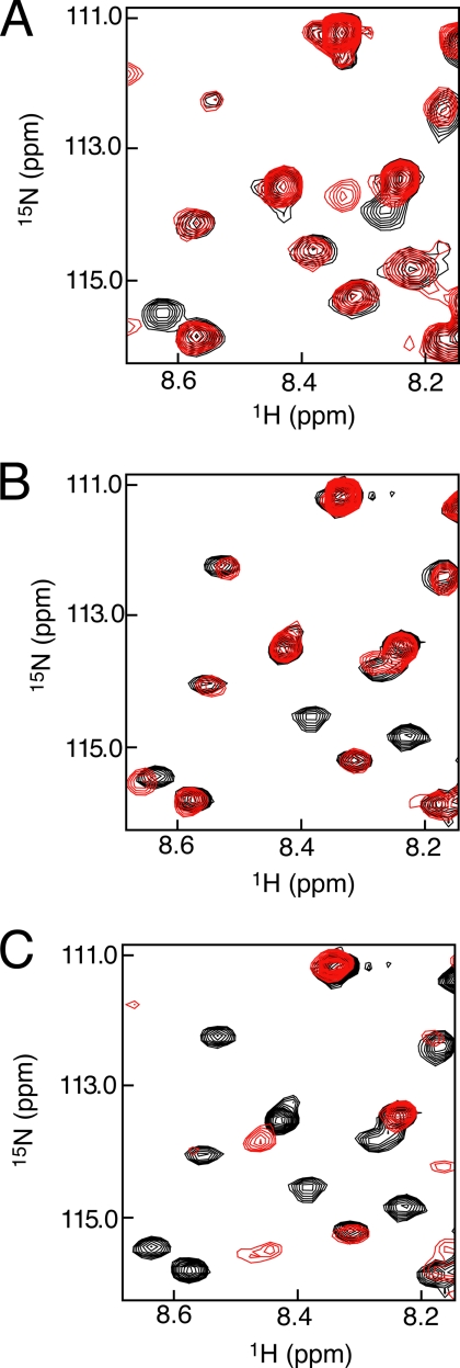
Inactivation of the retinoblastoma protein (Rb) through phosphorylation is an important step in promoting cell cycle progression, and hyperphosphorylated Rb is commonly found in tumors. Rb phosphorylation prevents its association with the E2F transcription factor; however, the molecular basis for complex inhibition has not been established. We identify here the key phosphorylation events and conformational changes that occur in Rb to inhibit the specific association between the E2F transactivation domain (E2F(TD)) and the Rb pocket domain. Calorimetry assays demonstrate that phosphorylation of Rb reduces the affinity of E2F(TD) binding approximately 250-fold and that phosphorylation at Ser(608)/Ser(612) and Thr(356)/Thr(373) is necessary and sufficient for this effect. An NMR assay identifies phosphorylation-driven conformational changes in Rb that directly inhibit E2F(TD) binding. We find that phosphorylation at Ser(608)/Ser(612) promotes an intramolecular association between a conserved sequence in the flexible pocket linker and the pocket domain of Rb that occludes the E2F(TD) binding site. We also find that phosphorylation of Thr(356)/Thr(373) inhibits E2F(TD) binding in a manner that requires the Rb N-terminal domain. Taken together, our results suggest two distinct mechanisms for how phosphorylation of Rb modulates association between E2F(TD) and the Rb pocket and describe for the first time a function for the structured N-terminal domain in Rb inactivation.


Figures

FIGURE 1.
FIGURE 1.
Domain structure of Rb and interactions with E2F-DP. A, Rb consists of a structured N-terminal domain (RbN) and central pocket domain. Its C-terminal domain (RbC) is disordered except for a short sequence that adopts a structure upon E2F binding. Two other unstructured sequences, the interdomain linker (RbIDL) and pocket linker (RbPL), are indicated. Structured regions are colored, and the conserved consensus Cdk phosphorylation sites are marked. B, Rb makes two distinct contacts with E2F. The pocket domain binds the E2F transactivation domain (E2FTD), and RbC binds the E2F-DP marked box domains.
FIGURE 2.
FIGURE 2.
Rb domain requirements for inhibition of E2FTD. A, ITC titration curves show that E2FTD binds to enzymatically dephosphorylated Rb55–928 (Rb55–928; Kd = 0.04 ± 0.02 μm) with a similar affinity as to the unphosphorylated Rb pocket domain (dephosRb380–787; Kd = 0.045 ± 0.007 μm). Phosphorylation of Rb55–928 results in a weaker affinity (phosRb55–928; Kd = 11 ± 3 μm). B, E2FTD dissociation constants were measured by ITC for binding to truncation mutants of Rb. The data demonstrate that RbN is required for phosphorylation-induced inhibition of E2FTD.
FIGURE 3.
FIGURE 3.
phosRbPL associates with the Rb pocket domain and competes with E2FTD binding. A, alignment of RbPL sequences from human (hs), mouse (mm), chicken (gg), frog (xl), and zebrafish (dr) shows that residues 595–611 (human) are highly conserved (yellow). B, HSQC spectra of 100 μm 15N-labeled phosRbPL592–624 alone (black) and in the presence of 500 μm unlabeled RbPΔPL (red). Broadening of amide resonances occurs selectively for residues 601–610, indicating a binding interaction between the phosphorylated pocket linker and pocket domain in trans. C, spectra of 100 μm 15N-labeled unphosphorylated RbPL alone (black) and in the presence of 400 μm unlabeled RbPΔPL (red). No resonance peak broadening is observed for unphosphorylated RbPL in the presence of the Rb pocket, demonstrating that binding is mediated by phosphorylation of RbPL. D, spectra of 100 μm 15N-labeled phosRbPL alone (black) and with 500 μm unlabeled RbPΔPL and 2 mm unlabeled E2FTD (red). In the presence of excess E2FTD, resonance peaks at chemical shifts corresponding to unbound phosRbPL reappear, indicating that E2FTD competes with phosRbPL for binding to the Rb pocket.
FIGURE 4.
FIGURE 4.
phosRbPL binds the pocket domain at the E2FTD binding site. A, structure of E2FTD bound to the pocket domain. Critical contacts between Asp424 and Phe426 of E2F and Arg467 and Phe482 of Rb are shown. This figure was generated using Protein Data Bank entry 1N4M. B and C, HSQC spectra of 100 μm 15N-labeled phosRbPL592–624 alone (black) and in the presence of 500 μm unlabeled RbPΔPL-R467A (red) and RbPΔPL-F482A (blue), respectively. D, resonance peak intensity ratios of phosRbPL in the presence of wild type RbPΔPL (black) and mutants R467A (red) and F482A (blue). The ratio I/I0 is defined as the peak intensity of phosRbPL in the presence of RbPΔPL (I) divided by the peak intensity of phosRbPL alone (I0). These data demonstrate that Arg467 and Phe482 in the pocket domain are critical for binding phosRbPL as well as E2FTD.
FIGURE 5.
FIGURE 5.
Detail of 1H-15N TROSY spectra of 300 μm2H-15N-labeled RbPΔPL alone (black) and in the presence (red) of unlabeled phosRbN-RbIDL (400 μm) (A), phosRbPL (1.5 mm) (B), and E2FTD (2 mm) (C). The observed spectral changes suggest that the binding sites for phosRbN-RbIDL and phosRbPL in the Rb pocket domain each partially overlap with the E2FTD binding site. Full spectra are shown in supplemental Fig. 3.

References

    1. Sherr C. J. (1996) Science 274, 1672–1677 - PubMed
    1. Weinberg R. A. (1995) Cell 81, 323–330 - PubMed
    1. Adams P. D. (2001) Biochim. Biophys. Acta 1471, M123–133 - PubMed
    1. Buchkovich K., Duffy L. A., Harlow E. (1989) Cell 58, 1097–1105 - PubMed
    1. Chen P. L., Scully P., Shew J. Y., Wang J. Y., Lee W. H. (1989) Cell 58, 1193–1198 - PubMed

Publication types

MeSH terms

LinkOut - more resources