. 2010 May 21;285(21):16231-8.
doi: 10.1074/jbc.M109.079236. Epub 2010 Mar 11.

Flightless-I (Fli-I) regulates the actin assembly activity of diaphanous-related formins (DRFs) Daam1 and mDia1 in cooperation with active Rho GTPase

Affiliations

Flightless-I (Fli-I) regulates the actin assembly activity of diaphanous-related formins (DRFs) Daam1 and mDia1 in cooperation with active Rho GTPase

Tomohito Higashi et al. J Biol Chem. .

Abstract

Eukaryotic cells dynamically reorganize the actin cytoskeleton to regulate various cellular activities, such as cell shape change, cell motility, cytokinesis, and vesicular transport. Diaphanous-related formins (DRFs), such as Daam1 and mDia1, play central roles in actin dynamics through assembling linear actin filaments. It has been reported that the GTP-bound active Rho binds directly to DRFs and partially unleashes the intramolecular autoinhibition of DRFs. However, whether proteins other than Rho involve the regulation of the actin assembly activity of DRFs has been unclear. Here, we show that Flightless-I (Fli-I), a gelsolin family protein essential for early development, binds directly to Daam1 and mDia1. Fli-I enhances the intrinsic actin assembly activity of Daam1 and mDia1 in vitro and is required for Daam1-induced actin assembly in living cells. Furthermore, Fli-I promotes the GTP-bound active Rho-mediated relief of the autoinhibition of Daam1 and mDia1. Thus, Fli-I is a novel positive regulator of Rho-induced linear actin assembly mediated by DRFs.


Figures

FIGURE 1.
FIGURE 1.
DAD segment enhances the actin assembly activity of Daam1 CT in cytoplasmic environment. A and B, domain structures of typical DRF (A) and Daam1 and mDia1 (B). The amino acid sequences of several DRF DAD segments are shown. GBD, GTPase-binding domain; DD, dimerization domain; Hs, Homo sapiens; Dm, Drosophila melanogaster; Ce, Caenorhabditis elegans; Sc, Saccharomyces cerevisiae. Asterisks in A show the mutated residues. C and D, cytoplasmic actin assembly assay (C) and pyrene actin assembly assay (D) of Daam1 CT mutants. The data shown in C are represented as means ± S.E. of five independent experiments, and the data shown in D are representative of three independent experiments with similar results. WT, wild type; LA, L1040A; FA, F1046A; a.u., arbitrary unit.
FIGURE 2.
FIGURE 2.
Fli-I binds to DAD segment of Daam1. A and B, Daam1 CT affinity chromatography. GST-Daam1 CT WT- or GST-Daam1 CT L1040A-coated beads were incubated with crude platelet cytosol or buffer alone, and the bound proteins were analyzed by Coomassie Brilliant Blue (CBB) staining (A) and immunoblot (IB) analysis using anti-Fli-I mAb (B). C, schematic representation of the domain structures of Fli-I and Gsn. LRR, leucine-rich repeat; G1–6, gelsolin motifs 1–6. D, direct binding of Daam1 to Fli-I. Beads coated with 50 pmol of GST, GST-Fli-I fragments, or GST-Gsn fragments were incubated with 62.5 nm His6-Daam1 CT. Bead-bound proteins were analyzed by SDS-PAGE and immunoblotting. E, specific interaction of Daam1 and Fli-I. Beads coated with 12.5 pmol of GST-Daam1 CT were incubated with 2.5 μm His6-Fli-I G4–6. Bead-bound proteins were analyzed by SDS-PAGE and immunoblotting. All the data shown in D and E are representative of three independent experiments with similar results.
FIGURE 3.
FIGURE 3.
Fli-I enhances the actin assembly activity of Daam1 CT in the cytoplasmic environment. A, immunodepletion of Fli-I. Human platelet cytosol was incubated with preimmune IgG- or anti-Fli-I IgG-coated beads. After centrifugation, the supernatant was analyzed by SDS-PAGE and immunoblotting with anti-Fli-I mAb and anti-actin mAb. Actin was used as a loading control. B, purified recombinant His6-tagged Fli-I was analyzed by SDS-PAGE followed by silver staining. C, cytoplasmic actin assembly was measured for 10 pmol of Daam1 CT in Fli-I-depleted cytosol in the absence or presence of 125 nm purified Fli-I. pre, preimmune IgG-treated cytosol; ID, Fli-I-immunodepleted cytosol. The data shown are represented as means ± S.E. of five independent experiments. a.u., arbitrary unit.
FIGURE 4.
FIGURE 4.
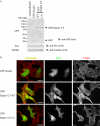
DAD segment of Daam1 enhances the actin assembly in living cells. A and B, NIH 3T3 cells were transiently transfected with GFP-Daam1 CT WT, GFP-Daam1 CT L1040A, or control GFP vector. After 20 h, the cell lysates were analyzed by SDS-PAGE and immunoblotting (IB) with anti-GFP, anti-Fli-I, and anti-tubulin mAbs (A), and cells were stained with Alexa-568-phalloidin (red). GFP signals were shown in green. WT, wild type; LA, L1040A. Scale bars, 20 μm (B). The data shown are representative of at least three independent experiments with similar results.
FIGURE 5.
FIGURE 5.
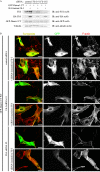
Fli-I is required for the actin assembly by Daam1 in living cells. A and B, NIH 3T3 cells were transfected with control or Fli-I siRNA. After 28 h, the cells were transiently transfected with GFP-Daam1 CT and/or hemagglutinin-tagged siRNA-insensitive Fli-I. After 20 h, cells were analyzed by SDS-PAGE and immunoblotting with anti-Fli-I, anti-GFP, anti-hemagglutinin, and anti-tubulin mAbs (A) or were stained with Alexa-568-phalloidin (red). GFP signals are shown in green. B, scale bars, 20 μm. The data shown are representative of at least three independent experiments with similar results.
FIGURE 6.
FIGURE 6.
Fli-I enhances the GTP-RhoA-mediated dissociation of the DID domain from the DAD segment. A, Fli-I competes with the DID domain on DAD binding. 50 pmol of GST-Daam1 DAD-coated beads were incubated with 25 pmol of His6-Fli-I G1–6 in the presence of various concentrations of His6-Daam1 NT. Bead-associated His6-Fli-I G1–6 was detected by immunoblotting (IB). B, concentration-dependent disruption of DID-DAD interaction by GTP-RhoA. Beads coated with 50 pmol of GST-Daam1 DAD were incubated with 125 nm His6-Daam1 NT in the presence of various concentrations of His6-RhoA G14V. Bead-associated His6-Daam1 NT was analyzed by immunoblotting with anti-His6 mAb. C, Fli-I enhances the DID-DAD dissociation of Daam1 by GTP-RhoA. Beads coated with 20 pmol of GST-Daam1 NT were incubated with 100 nm His6-Daam1 CT in the absence or presence of 0.1 μm His6-RhoA G14V and various concentrations of His6-Fli-I G4–6. Bead-bound His6-Daam1 CT was detected by immunoblotting. D, Fli-I promotes the Daam1 activation by GTP-RhoA. The actin nucleation activity of 50 nm Daam1 CT in the absence or presence of 200 nm Daam1 NT, 800 nm Fli-I G4–6, and 250 nm RhoA G14V was evaluated by the pyrene-actin assembly assay, as indicated. a.u., arbitrary unit. All the data shown in A–D are representative of 3–5 independent experiments with similar results.
FIGURE 7.
FIGURE 7.
Fli-I enhances the actin assembly activity of mDia1 as well as that of Daam1. A, cytoplasmic actin assembly assay of mDia1 CT mutants. WT, wild type; LA, L1197A; FA, F1203A. B, direct binding of mDia1 to Fli-I. Beads coated with 50 pmol of GST-tagged proteins were incubated with 12.5 pmol of His6-tagged proteins as indicated. Bound proteins were analyzed by SDS-PAGE and immunoblotting (IB). C, cytoplasmic actin assembly was measured for 1 pmol of mDia1 CT in Fli-I-depleted cytosol used in Fig. 3C in the absence or presence of 125 nm purified Fli-I. pre, preimmune IgG-treated cytosol; ID, Fli-I-immunodepleted cytosol. D, concentration-dependent disruption of DID-DAD interaction of mDia1 by GTP-RhoA. Beads coated with 50 pmol of GST-mDia1 DAD were incubated with 125 nm His6-mDia1 NT in the presence of various concentrations of His6-RhoA G14V. Bead-associated His6-mDia1 NT was analyzed by immunoblotting with anti-His6 mAb. E, Fli-I enhances the DID-DAD dissociation of mDia1 by GTP-RhoA. Beads coated with 100 nm GST-mDia1 NT were incubated with 20 pmol of His6-mDia1 CT in the absence or presence of 0.1 μm His6-RhoA G14V and various concentrations of His6-Fli-I G4–6. Bead-bound His6-mDia1 CT was detected by immunoblotting. F, Fli-I promotes the mDia1 activation by GTP-RhoA. The actin nucleation activity of 5 nm mDia1 CT in the absence or presence of 20 nm mDia1 NT, 400 nm Fli-I G4–6, and 100 nm RhoA G14V was evaluated by the pyrene-actin assembly assay, as indicated. The data shown in A and C are represented as means ± S.E. of five independent experiments, and the data shown in B and D–F are representative of three independent experiments with similar results. a.u., arbitrary unit.
FIGURE 8.
FIGURE 8.
Model for the regulation of DRF-mediated actin assembly by Rho and Fli-I. See text for details.

Similar articles

Cited by

References

    1. Faix J., Grosse R. (2006) Dev. Cell 10, 693–706 - PubMed
    1. Higgs H. N. (2005) Trends Biochem. Sci. 30, 342–353 - PubMed
    1. Paul A. S., Pollard T. D. (2009) Cell Motil. Cytoskeleton 66, 606–617 - PMC - PubMed
    1. Pruyne D., Evangelista M., Yang C., Bi E., Zigmond S., Bretscher A., Boone C. (2002) Science 297, 612–615 - PubMed
    1. Sagot I., Rodal A. A., Moseley J., Goode B. L., Pellman D. (2002) Nat. Cell Biol. 4, 626–631 - PubMed

Publication types

MeSH terms