

Accessory or additional renal arteries show no relevant effects on the width of the upper urinary tract: a 64-slice multidetector CT study in 1072 patients with 2132 kidneys
- PMID: 20223903
- PMCID: PMC3473859
- DOI: 10.1259/bjr/79479004
Accessory or additional renal arteries show no relevant effects on the width of the upper urinary tract: a 64-slice multidetector CT study in 1072 patients with 2132 kidneys
Abstract
Objective: The aim of this study was to find out on an unselected patient group whether crossing vessels have an influence on the width of the renal pelvis and what independent predictors of these target variables exist.
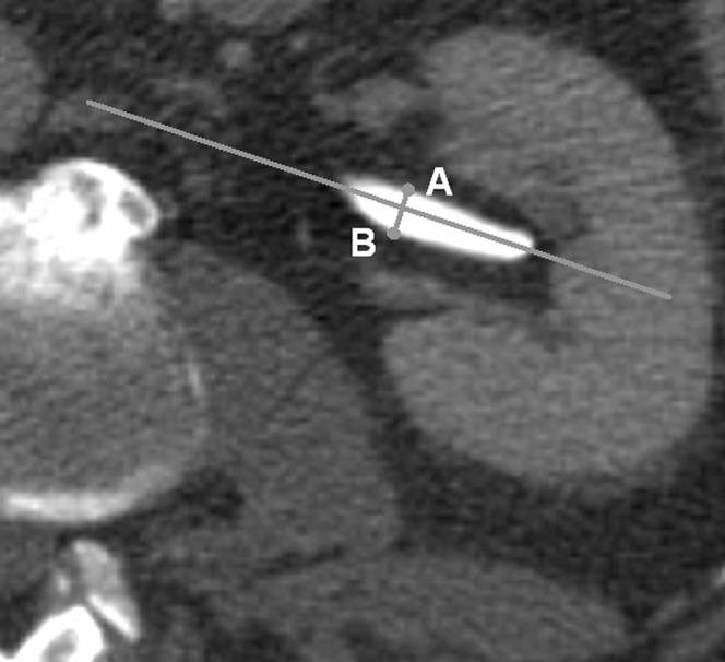
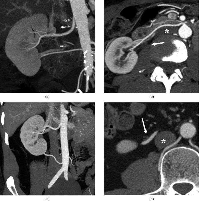
Methods: In this cross-sectional study, 1072 patients with arterially contrasted CT scans were included. The 2132 kidneys were supplied by 2736 arteries.
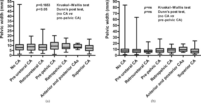
Results: On the right side, there were 293 additional and accessory arteries in 286 patients, and on the left side there were 304 in 271 patients. 154 renal pelves were more than 15 mm wide. The greatest independent factor for hydronephrosis on one side was hydronephrosis on the contralateral side (p<0.0001 each). Independent predictors for the width of the renal pelvis on the right side were the width of the renal pelvis on the left, female gender, increasing age and height; for the left side, predictors were the width of the renal pelvis on the right, concrements, parapelvic cysts and great rotation of the upper pole of the kidney to dorsal. Crossing vessels had no influence on the development of hydronephrosis. Only anterior crossing vessels on the right side are associated with widening of the renal pelvis by 1 mm, without making it possible to identify the vessel as an independent factor in multivariate regression models.
Conclusion: The width of the renal pelvis on the contralateral side is the strongest independent predictor for hydronephrosis and the width of the renal pelvis. There is no link between crossing vessels and the width of the renal pelvis.
Figures
Similar articles
-
Normal kidney size and its influencing factors - a 64-slice MDCT study of 1.040 asymptomatic patients.BMC Urol. 2009 Dec 23;9:19. doi: 10.1186/1471-2490-9-19. BMC Urol. 2009. PMID: 20030823 Free PMC article.
-
[Dodatkowa tętnica nerkowa przyczyną skrajnego wodonercza u 5-letniego chłopca].Pol Merkur Lekarski. 2018 Apr 23;44(262):201-204. Pol Merkur Lekarski. 2018. PMID: 29775449 Polish.
-
Precaval right renal arteries: prevalence and morphologic associations at spiral CT.Radiology. 2004 Feb;230(2):429-33. doi: 10.1148/radiol.2302021030. Radiology. 2004. PMID: 14752187
-
Evaluation of prospective living renal donors for laparoscopic nephrectomy with multisection CT: the marriage of minimally invasive imaging with minimally invasive surgery.Radiographics. 2001 Oct;21 Spec No:S223-36. doi: 10.1148/radiographics.21.suppl_1.g01oc10s223. Radiographics. 2001. PMID: 11598259 Review.
-
Computed tomography angiography with 3D reconstruction in diagnosis of hydronephrosis cause by aberrant renal vessel: A case report and mini review.J Xray Sci Technol. 2018;26(1):125-131. doi: 10.3233/XST-17343. J Xray Sci Technol. 2018. PMID: 29480234 Review.
Cited by
-
Laparoscopic transposition of lower pole crossing vessels (vascular hitch) in children with pelviureteric junction obstruction.Transl Pediatr. 2016 Oct;5(4):256-261. doi: 10.21037/tp.2016.09.08. Transl Pediatr. 2016. PMID: 27867849 Free PMC article.
-
Retrospective morphometric study of the suitability of renal arteries for renal denervation according to the Symplicity HTN2 trial criteria.BMJ Open. 2016 Jan 4;6(1):e009351. doi: 10.1136/bmjopen-2015-009351. BMJ Open. 2016. PMID: 26729385 Free PMC article.
References
-
- Johnston JH. The pathogenesis of hydronephrosis in children. Br J Urol 1969;41:724–34 - PubMed
-
- Zhang X, Xu K, Fu B, Zhang J, Lang B, Ai X, et al. The retroperitoneal laparoscopic Hellström technique for pelvi-ureteric junction obstruction from a crossing vessel. BJU Int 2007;100:1335–8 - PubMed
-
- Braun P, Guilabert JP, Kazmi F. Multidetector computed tomography arteriography in the preoperative assessment of patients with ureteropelvic junction obstruction. Eur J Radiol 2007;61:170–5 - PubMed
-
- Sampaio FJ. Vascular anatomy at the ureteropelvic junction. Urol Clin North Am 1998;25:251–8 - PubMed
-
- Rehman J, Landman J, Sundaram C, Clayman RV. Missed anterior crossing vessels during open retroperitoneal pyeloplasty: laparoscopic transperitoneal discovery and repair. J Urol 2001;166:593–6 - PubMed
