

Longitudinal MRI shows no cerebral abnormality in chronic fatigue syndrome
- PMID: 20223910
- PMCID: PMC3473570
- DOI: 10.1259/bjr/85621779
Longitudinal MRI shows no cerebral abnormality in chronic fatigue syndrome
Abstract
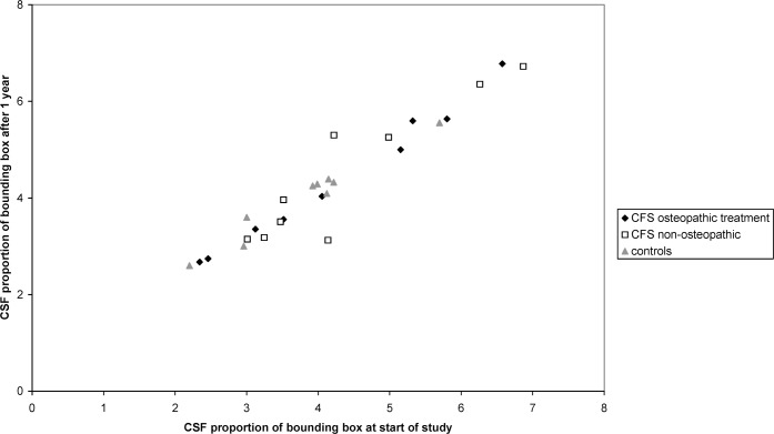
MRI has previously provided conflicting results when used to search for brain abnormalities in sufferers of chronic fatigue syndrome (CFS). Eighteen CFS patients and nine healthy volunteers each underwent MRI on two occasions, one year apart. The resulting images were examined for abnormalities in brain atrophy, deep white matter hyperintensities (WMH) and cerebral blood and cerebrospinal fluid (CSF) flow. Mean proportionate CSF volume was not significantly different between subject groups. All participants showed a slight increase in CSF between scans, but no significant difference was found between those with CFS and those without. Between-group comparisons of ventricular volume revealed no significant differences at study commencement and no significant change over the year. No significant inter-group differences were found for any of the cerebral blood and CSF flow parameters. Low levels of WMH were found in all participants. Objective scoring of WMH using Scheltens' scale revealed no change in summary components (prosencephalic deep white matter hyperintensities, basal ganglia hyperintensities and infratentorial hyperintensities) or in individual component variables between the baseline and 1 year follow-up scans. No abnormal patterns in rate and extent of brain atrophy, ventricle volume, white matter lesions, cerebral blood flow or aqueductal CSF flow were detected in the CFS population. These results throw open the debate into whether MRI scanning can reveal diagnostic signs of CFS and clinically questions the diagnoses of CFS made on the basis of previous research conclusions.
Figures
References
-
- Lange G, DeLuca J, Maldjian JA, Lee H, Tiersky LA, Natelson BH. Brain MRI abnormalities exist in a subset of patients with chronic fatigue syndrome. J Neurol Sci 1999;171:3–7 - PubMed
-
- Lange G, Holodny AI, DeLuca J, Huey-Jen L, Xiao-Hong MY, Steffener J, Natelson BH. Quantitative assessment of cerebral ventricular volumes in chronic fatigue syndrome. Appl Neuropsychol 2001;8:23–30 - PubMed
-
- Cope H, Pernet A, Kendall B, David A. Cognitive functioning and magnetic resonance imaging in chronic fatigue. Br J Psychiatry 1995;167:86–94 - PubMed
-
- Schwartz RB, Garada BM, Komaroff AL, Tice HM, Gleit M, Jolesz FA, Holman BL. Detection of intracranial abnormalities in patients with chronic fatigue syndrome: comparison of MR imaging and SPECT. AJR Am J Roentgenol 1994;162:935–41 - PubMed
Publication types
MeSH terms
LinkOut - more resources
Full Text Sources
Medical
