

doi: 10.1126/science.1184008.
Epub 2010 Mar 11.
A peroxidase/dual oxidase system modulates midgut epithelial immunity in Anopheles gambiae
Affiliations
- PMID: 20223948
- PMCID: PMC3510679
- DOI: 10.1126/science.1184008
Item in Clipboard
A peroxidase/dual oxidase system modulates midgut epithelial immunity in Anopheles gambiae
Science.
.
Abstract
Extracellular matrices in diverse biological systems are cross-linked by dityrosine covalent bonds catalyzed by the peroxidase/oxidase system. We show that a peroxidase, secreted by the Anopheles gambiae midgut, and dual oxidase form a dityrosine network that decreases gut permeability to immune elicitors. This network protects the microbiota by preventing activation of epithelial immunity. It also provides a suitable environment for malaria parasites to develop within the midgut lumen without inducing nitric oxide synthase expression. Disruption of this barrier results in strong and effective pathogen-specific immune responses.
Figures
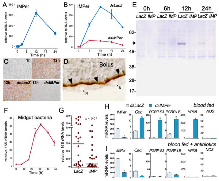
Expression and localization of IMPer and effect of IMPer silencing on bacterial growth and immune activation. (A) IMPer mRNA expression and (B) effect of IMPer silencing on IMPer mRNA levels at different times after blood feeding (mean ± SEM). (C) Effect of IMPer silencing on midgut peroxidase activity detected using 3,3′-diaminobenzidine (DAB) staining at different times after feeding. (D) Localization of inducible peroxidase activity detected using DAB staining (brown) in midgut sections 12 h after feeding. N, nucleus of epithelial cells. (E) Western blot of IMPer protein in midgut homogenates collected at different times after feeding in control (dsLacZ injected) or IMPer-silenced mosquitoes. (F) Bacterial 16S rRNA levels in midguts collected at different times after blood feeding (mean ± SEM). (G) Effect of IMPer silencing on bacterial 16S rRNA of individual midguts 24 h after feeding (line indicates the median) (mean ± SEM). (H) IMPer, Cecropin, PGRP-S3, PGRP-LB, HPX8, and NOS mRNA levels in control (dsLacZ-injected) and IMPer-silenced midguts 24 h after feeding (mean ± SEM). (I) Same as (H) but in antibiotic-fed mosquitoes.
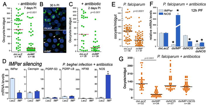
Effect of IMPer silencing on Plasmodium infection and midgut immune activation. Effect of IMPer silencing on P. berghei infection (A) 7 days post infection (PI), (B) 30 h PI, and (C) 2 days PI in antibiotic-fed mosquitoes. (D) IMPer, Cecropin, PGRP-S3, PGRP-LB, HPX8, and NOS mRNA levels in control (dsLacZ-injected) and IMPer-silenced midguts of antibiotic-treated mosquitoes 24 h PI (mean ± SEM). (E) Effect of IMPer silencing on P. falciparum infection 8 days PI. (F) Effect of silencing IMPer, NOS, or co-silencing IMPer and NOS, on IMPer and NOS mRNA expression 12 h PI in antibiotic-fed mosquitoes infected with P. falciparum (mean ± SEM) and (G) P. falciparum infection 7 days PI. Asterisks indicate significant differences relative to the dsLacZ control. Each circle represents the number of parasites in an individual midgut, and the line indicates the median.
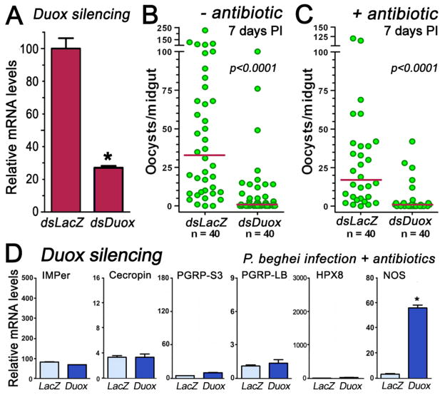
Effect of Duox silencing on Plasmodium infection and midgut immune activation. (A) Midgut Duox silencing 4 days post injection of dsDoux (B) Effect of Duox silencing on P. berghei infection 7 days post infection (PI). (C) Same as (B), but in antibiotic-fed females. (D) Effect of Duox silencing on Cecropin, PGRP-S3, PGRP-LB, heme-peroxidase 8, and NOS expression in midguts of antibiotic-treated mosquitoes collected 24 h PI (mean ± SEM).
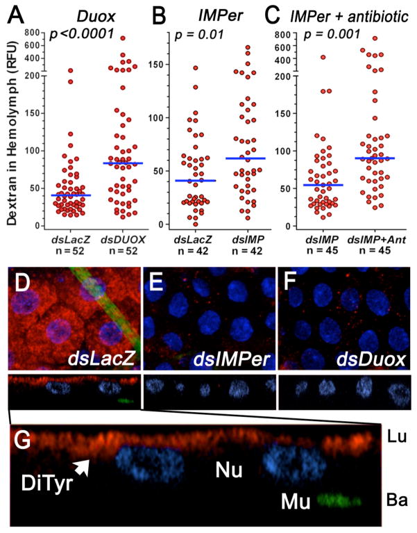
Gut permeability and dityrosine network. Effect of (A) Duox or (B) IMPer silencing without or (C) with oral antibiotics (Ant.) on midgut permeability to fluorescent dextran (4 kDa). Each circle represents fluorescence in the hemolymph of an individual mosquito 18–20 h after feeding (line indicates the median). (D–G) Immunofluorescence staining of midguts 14 h after feeding. Dityrosine bonds (red) and muscle actin (green) in mosquitoes injected with (D) dsLacZ, (E) dsIMPer, or (F) dsDuox. (G) Enlargement of (D). DiTyr, dityrosine staining; Lu, lumen; Ba, basal; Mu, muscle; Nu, nuclei.
References
Publication types
MeSH terms
Substances
Associated data
- Actions
Grants and funding
LinkOut - more resources
Full Text Sources
Molecular Biology Databases
