. 2010 Mar 30;107(13):6034-9.
doi: 10.1073/pnas.0914718107. Epub 2010 Mar 11.

Zebrafish larvae lose vision at night

Affiliations

Zebrafish larvae lose vision at night

Farida Emran et al. Proc Natl Acad Sci U S A. .

Abstract

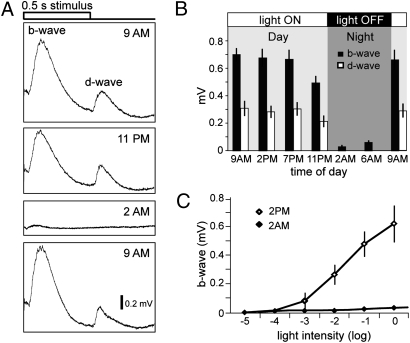
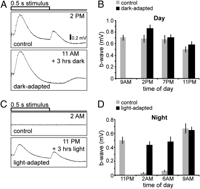
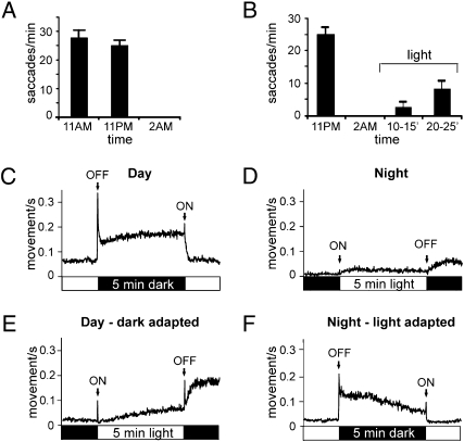
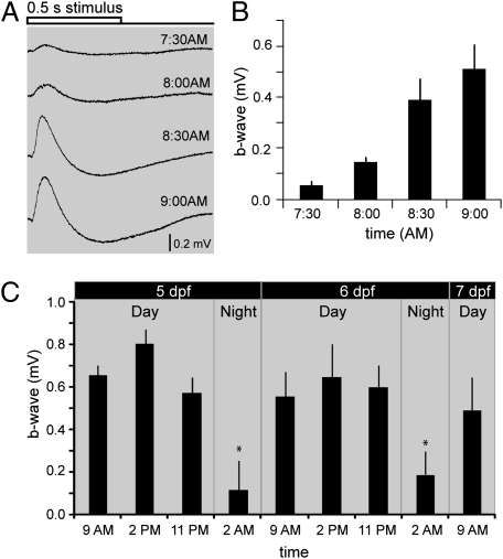
Darkness serves as a stimulus for vertebrate photoreceptors; they are actively depolarized in the dark and hyperpolarize in the light. Here, we show that larval zebrafish essentially turn off their visual system at night when they are not active. Electroretinograms recorded from larval zebrafish show large differences between day and night; the responses are normal in amplitude throughout the day but are almost absent after several hours of darkness at night. Behavioral testing also shows that larval zebrafish become unresponsive to visual stimuli at night. This phenomenon is largely circadian driven as fish show similar dramatic changes in visual responsiveness when maintained in continuous darkness, although light exposure at night partially restores the responses. Visual responsiveness is decreased at night by at least two mechanisms: photoreceptor outer segment activity decreases and synaptic ribbons in cone pedicles disassemble.


Conflict of interest statement

The authors declare no conflict of interest.

Figures

Fig. 1.
Fig. 1.
Larval zebrafish exhibit a day/night cycle of visual responsiveness. (A) Representative ERG recordings to a 0.5 second white light stimulus are shown at four different time points: at 9:00 AM, when the ambient lights are turned ON; 11:00 PM, shortly before the lights are turned OFF; 2:00 AM, after the lights have been OFF for 3 h; and at 9:00 AM the next morning, after the lights are turned ON again. Note the large reduction of ERG amplitudes at 2:00 AM. (B) Average ERG b- (black bars) and d-wave (white bars) amplitudes taken at different time points over a 24-h period (n = 15 fish for each time point; bars indicate SD). (C) V-log I curves showing that increasing the light stimulus intensity at 2:00 AM did not increase the ERG amplitudes significantly. Average b-wave amplitudes in response to 5 log units of white light stimulus intensities at 2:00 PM and 2:00 AM (n = 5 fish for each time point).
Fig. 2.
Fig. 2.
Exposure to light inhibits the loss of visual responsiveness at night. (A) Dark-adaptation during the day causes a slight increase in b-wave amplitude. Representative ERG recordings are shown for animals exposed to normal light conditions (control) and for animals exposed to 3 h of darkness during the day (dark-adapted). (B) Comparison of average b-wave amplitudes for control fish (gray bars) and dark-adapted fish (black bars) taken at different time points throughout the day (n = 15 fish for each time point; averaged b-wave amplitudes are compared to their respective control; bars indicate SD). (C) Light-adaptation at night partially overrides the loss of visual responsiveness and prevents the severe decrease of b- and d-wave amplitudes. Representative ERGs for control animals (Upper) and for animals exposed to 3 h of light (Lower) recorded at 2:00 AM. (D) Light-adapted b-wave amplitudes during the night increased as dawn approaches and are similar to control b-wave amplitudes by 9:00 AM when the lights are turned ON. Comparison of averaged b-wave amplitudes for control fish (gray bars) and fish exposed to light (black bars) throughout the night starting at 11:00 PM taken at different time points (n = 15 fish for each time point; averaged b-wave amplitudes are compared with their respective control; bars indicate SD).
Fig. 3.
Fig. 3.
Zebrafish behavioral responses to light are abolished at night but can be partially restored by continuous exposure to light. (A) Larval zebrafish exhibit a robust OKR at 11:00 AM and 11:00 PM (shortly before the lights are turned OFF at night) but not at 2 AM (3 h after the lights were turned OFF, n = 20 for each time point). (B) Light-adaptation during the night reverses the loss of OKR responses. OKR responses were taken at 11 PM, 2 AM, and at two time points after exposure to light starting at 2 AM: 10–15 and 20–25 min. (C) Larval zebrafish displayed ON and OFF visual-motor responses VMR during the day but fail to exhibit transient startle responses at night (each trace represents an average of 240 responses from 80 individual fish). During the daytime, motor output increased in response to a 5-min light off stimulus with prominent OFF and ON responses (first stimulus was introduced at 2:00 PM). (D) At night, the VMR responses to a 5-min light stimulus were abolished (first stimulus was introduced at 2:00 AM). (E) Dark-adaptation during the day does not abolish VMR responses. Fish, dark-adapted for 3 h, were then exposed to a 5-min light stimulus (first light stimulus was introduced at 2:00 PM). (F) Light-adaptation at night prevents the loss of visual responsiveness and restores VMR responses. Fish were first light-adapted for 3 h starting at 11:00 PM and then exposed to 5-min of darkness (first lights off stimulus was introduced at 2:00 AM).
Fig. 4.
Fig. 4.
Loss and recovery of visual responsiveness occurs within tens of minutes. (A) Visual responsiveness gradually decreases over 1 h of darkness and is abolished after 90 min of darkness at night. Representative ERG traces are shown for 11:00 PM, recorded right before the ambient light is turned OFF at night, and at different time points after the lights are turned OFF. (B) Averaged b-wave amplitudes at different time points (n = 5 per time point) after the lights are turned OFF at night. (C) Visual responsiveness is partially restored within 35 min after the lights are turned ON during the night. Representative ERGs are shown for 2:00 AM at night, and at different time points after the lights are turned ON. (D) Averaged b-wave amplitudes at different time points (n = 5 per time points) after the lights are turned ON at 2:00 AM.
Fig. 5.
Fig. 5.
A circadian clock regulates visual responsiveness in larval zebrafish. (A) Zebrafish larvae anticipate sunrise and exhibit an increase in b-wave amplitudes before the lights are turned ON (at 9:00 AM). Representative ERG recordings taken at different time points before the lights are turned ON in the morning. (B) Averaged b-wave amplitudes at different time points (n = 5 per time point) before the lights are turned ON in the morning. c) Visual responsiveness is regulated by a circadian cycle. Averaged b-wave amplitudes at different time points (n ≥ 10) over 48 h of darkness starting at day 5 before the lights were turned on. A consistent cycle of visual responsiveness is observed: b-wave amplitudes are high during the day (9:00 AM, 2:00 PM, 11:00 PM), become significantly smaller at night (2:00 AM), but are increased by the next morning (9:00 AM) for several days.
Fig. 6.
Fig. 6.
Photoreceptor function is altered at night. (A) ERG recordings from eyes of fish incubated in fish water (control) and in fish water containing a drug mixture to block the b-wave. The a-wave, reflecting photoreceptor outer segment hyperpolarization and revealed after drug treatment, was significantly smaller at night (2 AM) than during the day (2:00 PM). (B) Averaged a-wave amplitudes (n = 15) taken from drug-treated fish at a 12-h interval (2:00 PM and 2:00 AM); and from fish that were exposed to light for 3 h at night. A-wave amplitudes at 2 AM were reduced by about 65% from those measured at 2:00 PM. Three hours of light adaptation at night increased a-wave amplitudes to control level (*, P < 0.01 and bars indicate SD). (C) EM sections of cone pedicles at different times of the day and under different exposures to light. Most cone pedicles exhibit prominent synaptic ribbons during the day (2:00 PM), but are essentially devoid of synaptic ribbons at night (2:00 AM). When fish are exposed to darkness for 3 h during the day (dark-adapted 2:00 PM) synaptic ribbons are still observed. Exposure to 3-h day light conditions at night (light-adapted 2:00 AM) prevents the degradation of synaptic ribbons that usually occurs at night and synaptic ribbons are observed. Arrows denote synaptic ribbons.

References

    1. Tomita T. Electrophysiological study of the mechanisms subserving color coding in the fish retina. Cold Spring Harb Symp Quant Biol. 1965;30:559–566. - PubMed
    1. Hagins WA, Penn RD, Yoshikami S. Dark current and photocurrent in retinal rods. Biophys J. 1970;10:380–412. - PMC - PubMed
    1. Tomita T. Electrical activity of vertebrate photoreceptors. Q Rev Biophys. 1970;3:179–222. - PubMed
    1. Wangsa-Wirawan ND, Linsenmeier RA. Retinal oxygen: Fundamental and clinical aspects. Arch Ophthalmol. 2003;121:547–557. - PubMed
    1. Okawa H, Sampath AP, Laughlin SB, Fain GL. ATP consumption by mammalian rod photoreceptors in darkness and in light. Curr Biol. 2008;18:1917–1921. - PMC - PubMed

Publication types

LinkOut - more resources