

Structural determinants in phycotoxins and AChBP conferring high affinity binding and nicotinic AChR antagonism
- PMID: 20224036
- PMCID: PMC2851920
- DOI: 10.1073/pnas.0912372107
Structural determinants in phycotoxins and AChBP conferring high affinity binding and nicotinic AChR antagonism
Abstract
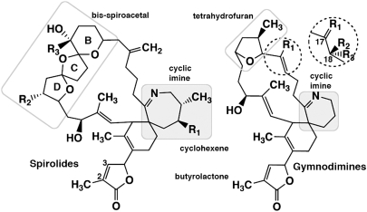
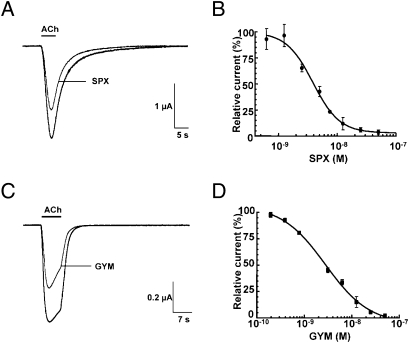
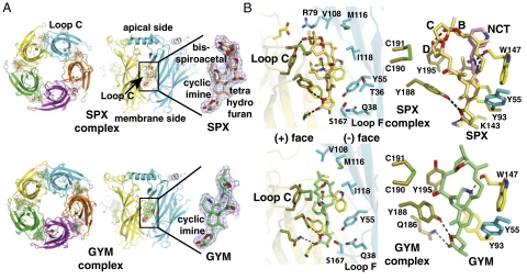
Spirolide and gymnodimine macrocyclic imine phycotoxins belong to an emerging class of chemical agents associated with marine algal blooms and shellfish toxicity. Analysis of 13-desmethyl spirolide C and gymnodimine A by binding and voltage-clamp recordings on muscle-type alpha1(2)betagammadelta and neuronal alpha3beta2 and alpha4beta2 nicotinic acetylcholine receptors reveals subnanomolar affinities, potent antagonism, and limited subtype selectivity. Their binding to acetylcholine-binding proteins (AChBP), as soluble receptor surrogates, exhibits picomolar affinities governed by diffusion-limited association and slow dissociation, accounting for apparent irreversibility. Crystal structures of the phycotoxins bound to Aplysia-AChBP ( approximately 2.4A) show toxins neatly imbedded within the nest of ar-omatic side chains contributed by loops C and F on opposing faces of the subunit interface, and which in physiological conditions accommodates acetylcholine. The structures also point to three major features: (i) the sequence-conserved loop C envelops the bound toxins to maximize surface complementarity; (ii) hydrogen bonding of the protonated imine nitrogen in the toxins with the carbonyl oxygen of loop C Trp147 tethers the toxin core centered within the pocket; and (iii) the spirolide bis-spiroacetal or gymnodimine tetrahydrofuran and their common cyclohexene-butyrolactone further anchor the toxins in apical and membrane directions, along the subunit interface. In contrast, the se-quence-variable loop F only sparingly contributes contact points to preserve the broad receptor subtype recognition unique to phycotoxins compared with other nicotinic antagonists. These data offer unique means for detecting spiroimine toxins in shellfish and identify distinctive ligands, functional determinants and binding regions for the design of new drugs able to target several receptor subtypes with high affinity.
Conflict of interest statement
The authors declare no conflict of interest.
Figures
References
-
- Ciminiello P, Fattorusso E. Bivalve molluscs as vectors of marine biotoxins involved in seafood poisoning. Prog Mol Subcell Biol. 2006;43:53–82. - PubMed
-
- Gill S, et al. Neural injury biomarkers of novel shellfish toxins, spirolides: A pilot study using immunochemical and transcriptional analysis. Neurotoxicology. 2003;24:593–604. - PubMed
-
- O’Connor PD, Brimble MA. Synthesis of macrocyclic shellfish toxins containing spiroimine moieties. Nat Prod Rep. 2007;24:869–885. - PubMed
-
- Cembella AD, Lewis NI, Quilliam MA. Spirolide composition of micro-extracted pooled cells isolated from natural plankton assemblages and from cultures of the dinoflagellate Alexandrium ostenfeldii. Nat Toxins. 1999;7:197–206. - PubMed
-
- Hu T, et al. Characterization of spirolides a, c, and 13-desmethyl c, new marine toxins isolated from toxic plankton and contaminated shellfish. J Nat Prod. 2001;64:308–312. - PubMed
Publication types
MeSH terms
Substances
Associated data
- Actions
- Actions
Grants and funding
LinkOut - more resources
Full Text Sources
Other Literature Sources
Molecular Biology Databases
Research Materials
Miscellaneous
