. 2010 Apr 30;106(8):1394-403.
doi: 10.1161/CIRCRESAHA.109.210286. Epub 2010 Mar 11.

Matrix-specific protein kinase A signaling regulates p21-activated kinase activation by flow in endothelial cells

Affiliations

Matrix-specific protein kinase A signaling regulates p21-activated kinase activation by flow in endothelial cells

Steven Daniel Funk et al. Circ Res. .

Abstract

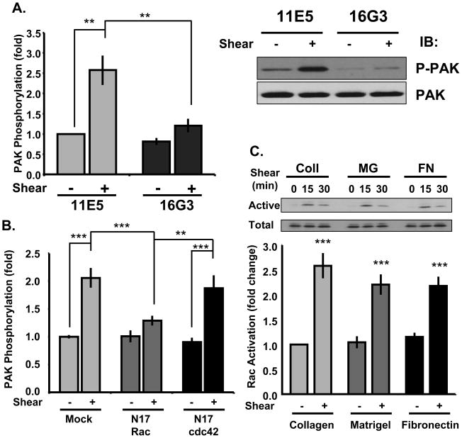
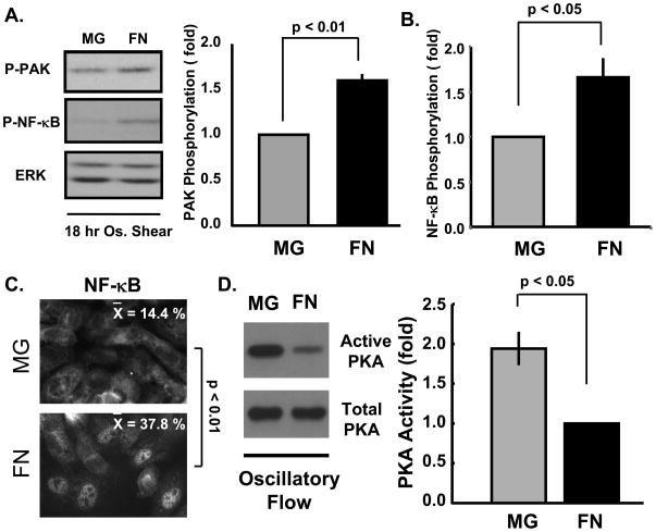
Rationale: Atherosclerosis is initiated by blood flow patterns that activate inflammatory pathways in endothelial cells. Activation of inflammatory signaling by fluid shear stress is highly dependent on the composition of the subendothelial extracellular matrix. The basement membrane proteins laminin and collagen found in normal vessels suppress flow-induced p21 activated kinase (PAK) and nuclear factor (NF)-kappaB activation. By contrast, the provisional matrix proteins fibronectin and fibrinogen found in wounded or inflamed vessels support flow-induced PAK and NF-kappaB activation. PAK mediates both flow-induced permeability and matrix-specific activation of NF-kappaB.

Objective: To elucidate the mechanisms regulating matrix-specific PAK activation.

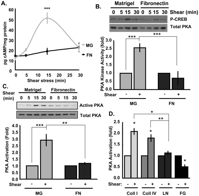
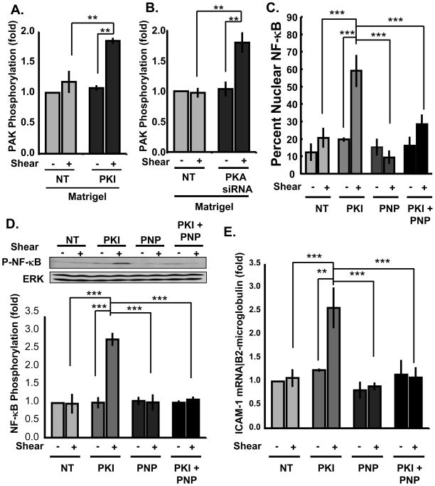
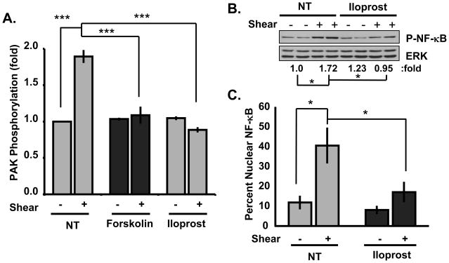
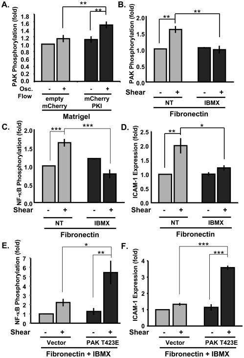
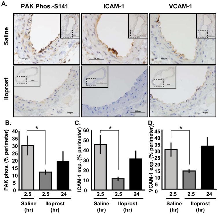
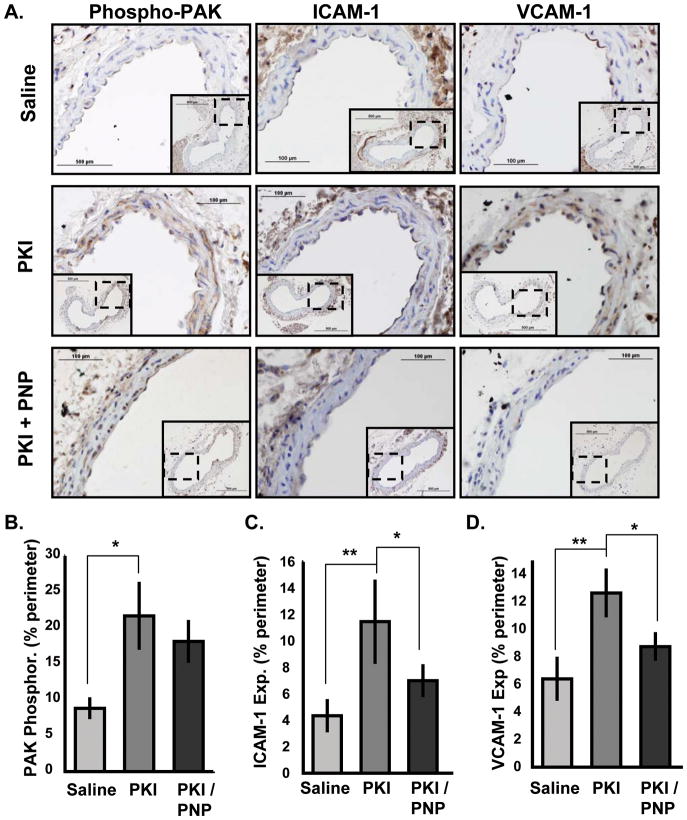
Methods and results: We now show that matrix composition does not affect the upstream pathway by which flow activates PAK (integrin activation, Rac). Instead, basement membrane proteins enhance flow-induced protein kinase (PK)A activation, which suppresses PAK. Inhibiting PKA restored flow-induced PAK and NF-kappaB activation in cells on basement membrane proteins, whereas stimulating PKA inhibited flow-induced activation of inflammatory signaling in cells on fibronectin. PKA suppressed inflammatory signaling through PAK inhibition. Activating PKA by injection of the prostacyclin analog iloprost reduced PAK activation and inflammatory gene expression at sites of disturbed flow in vivo, whereas inhibiting PKA by PKA inhibitor (PKI) injection enhanced PAK activation and inflammatory gene expression. Inhibiting PAK prevented the enhancement of inflammatory gene expression by PKI.

Conclusions: Basement membrane proteins inhibit inflammatory signaling in endothelial cells via PKA-dependent inhibition of PAK.


Figures

Figure 1
Figure 1. Analysis of the PAK upstream pathway
(A) BAECs on fibronectin were treated with fibronectin-blocking (16G3, 50 μg/ml) or nonblocking (11E5, 50 μg/ml) antibodies for 30 minutes. Cells were sheared for 15 minutes and PAK phosphorylation was determined by Western blotting for phospho-Ser141. Results are normalized to total protein. n = 3. (B) ECs expressing N17-Rac or N17-Cdc42 plated on a fibronectin matrix were sheared for 15 minutes. PAK activation was determined as described. n = 3–5. (C) ECs plated on collagen (Coll), matrigel (MG), or fibronectin (FN) were sheared for the indicated times and Rac activity was determined by affinity pulldown assays. Rac levels in the pulldown at the 0 and 15 minute time points were normalized to total Rac levels in whole cell lysates. n = 4–6. ** p < 0.01; *** p < 0.001 by multiple comparisons ANOVA.
Figure 2
Figure 2. Matrix-specific PKA activation
BAECs plated on matrigel (MG) or fibronectin (FN)-coated slides were exposed to onset of laminar flow for the indicated times. (A) cAMP concentration was determined by competitive ELISA assay and normalized to total protein. n = 4–6. (B) Kinase activity in PKA immunoprecipitates was determined in a kinase assay using GST-CREB as substrate and antibodies specific for phosphorylated CREB. Results at the 0 and 15 minute time points were normalized to PKA levels in the immunoprecipitates and for total CREB. n = 3. (C) PKA activity was determined by affinity pulldown using GST-PKI. Active PKA in the pulldowns was normalized to total PKA in cell lysates using the 0 and 15 min. time points. n = 3. (D) ECs plated on slides coated with collagen I (Coll I), collagen IV (Coll IV), laminin (LN), or fibrinogen (FG) were sheared for 15 minutes, and PKA activity determined by GST-PKI pulldown. n = 3–5. * p < 0.05, ** p < 0.01, *** p < 0.001 by multiple comparisons ANOVA.
Figure 3
Figure 3. PKA inhibitors rescue inflammatory signaling on MG
(A) BAECs plated on matrigel were treated with the cell permeant PKA inhibitory peptide PKI (20 μM for 15 min.). Cells were then sheared for 15 minutes and PAK assayed as in Fig 1. n = 3. (B) ECs transfected with siRNA against the PKC Cα catalytic subunit (~70% knockdown) were plated on matrigel and sheared for 15 min. PAK was assayed as above. n = 3. (C,D) ECs plated on matrigel were treated with PKI (20 μM for 15 min.), the PAK inhibitory peptide (PNP; 20 μg/ml for 60 min.) or both. Cells were then sheared for 30 minutes and NF-κB nuclear translocation (C) and phosphorylation on Ser536 (D) were determined. Western blots were normalized to total protein, and representative blots are shown. n = 3. For nuclear translocation, more than 100 cells were counted for each condition and scored as positive or negative for nuclear NF-κB. NF-κB nuclear translocation was averaged from three independent experiments. (E) HAECs were plated on matrigel were treated with PKI (20 μM for 15 min.), the PAK inhibitory peptide (PNP; 20 μg/ml for 60 min.) or both and then sheared for 3 hours. ICAM-1 mRNA expression was determined using quantitative real time PCR and normalized toβ2-microglobulin mRNA. ** p < 0.01, *** p < 0.001 by multiple comparisons ANOVA.
Figure 4
Figure 4. PKA activators inhibit inflammatory signaling on fibronectin
(A) BAECs plated on fibronectin were treated with forskolin (10 μM) or iloprost (1 μM) for 30 minutes. Cells were sheared for 15 minutes and PAK was assayed as in Figure 1. n = 3–6. (B,C) ECs plated on fibronectin were treated with iloprost (1 μM for 30 min.), sheared for 30 min., and (B) NF-κB phosphorylation on Ser536 and (C) nuclear translocation were determined as in Fig 3. n = 3. * p < 0.05, *** p < 0.001 by multiple comparisons ANOVA.
Figure 5
Figure 5. Matrix-specific signaling in oscillatory flow
BAECs plated on matrigel or fibronectin were exposed to oscillatory flow for 18 hours. Activation of (A) PAK and (A,B) NF-κB were determined by Western blotting with phospho-specific antibodies as previously described. n = 3–4. (C) Cells were fixed and NF-κB nuclear translocation was determined as in previous figures. Representative images are shown. n = 3 (D) PKA activity was determined using the GST-PKI affinity beads. Active PKA in the pulldown was normalized to total PKA in the lysate. n = 3. Statistical method was student’s T-test.
Figure 6
Figure 6. PAK mediates anti-inflammatory PKA signaling in oscillatory flow
(A) BAECs transfected (50% efficiency) with control, empty mCherry vector or mCherry-PKI were plated on matrigel and exposed to oscillatory flow for 18 hours. PAK activation was determined as in Fig 1. n = 4. (B) HAECs plated on fibronectin were exposed to oscillatory flow for 18 hours in the presence of the PDE inhibitor IBMX (100 μM) or vehicle control. (B) PAK phosphorylation and (C) NF-κB phosphorylation were determined. (D) ICAM-1 expression was determined by Western blotting and normalized to total protein. n = 3. (E,F) HAECs transfected with Myc-tagged T423E-PAK or control vector were plated on fibronectin and exposed to oscillatory flow in the presence of IBMX (100 μM) for 18 hours. (E) NF-κB phosphorylation and (F) ICAM-1 expression were determined by Western blotting as previously described. n = 3. * p < 0.05, ** p < 0.01 by multiple comparisons ANOVA.
Figure 7
Figure 7. Iloprost reduces PAK activation in areas of disturbed flow in vivo
(A) C57Bl/6J mice at 36–38 weeks received intraperitoneal injection of iloprost (20 μg; ~1 mg/kg) or saline control. After 2.5 or 24 hours, carotid arteries were collected and analyzed for PAK phosphorylation (Ser141), ICAM-1 expression, and VCAM-1 expression by immunohistochemistry. 40X images show the atheroprone region at the outer wall of the carotid sinus. 10X images show the entire artery (insets). (B,C,D) The percent of the vessel lumen staining positive for (B) phosphorylated PAK, (C) ICAM-1, and (D) VCAM-1 was determined. n = 3. * p < 0.05 by one-way ANOVA.
Figure 8
Figure 8. PKA inhibition stimulates PAK activation and inflammation in vivo
(A) C57Bl/6J mice at 10 weeks old received retroorbital injection of saline, PKI (24 μg, ~1.2 mg/kg) or PKI and the PAK inhibitory peptide (PNP, 50 μg, ~2.5 mg/kg). After 2.5 hours, carotid arteries were collected and analyzed for PAK phosphorylation (Ser141), ICAM-1 expression, and VCAM-1 expression by immunohistochemistry. 40X images show the atheroprone region at the outer wall of the carotid sinus. 10X images show the entire artery (insets). (B,C,D) The percent of the vessel lumen staining positive for (B) phosphorylated PAK, (C) ICAM-1, and (D) VCAM-1 was determined. n = 3. * p < 0.05 by one-way ANOVA.

Similar articles

Cited by

References

    1. Ross R. Atherosclerosis--an inflammatory disease. New England Journal of Medicine. 1999;340:115–126. [see comment] - PubMed
    1. Gimbrone MA, Jr, Topper JN, Nagel T, Anderson KR, Garcia-Cardena G. Endothelial dysfunction, hemodynamic forces, and atherogenesis. Annals of the New York Academy of Sciences. 2000;902:230–239. discussion 239–240. - PubMed
    1. Hahn C, Schwartz MA. Mechanotransduction in vascular physiology and atherogenesis. Nat Rev Mol Cell Biol. 2009;10:53–62. - PMC - PubMed
    1. Mohan S, Mohan N, Sprague EA. Differential activation of NF-kappa B in human aortic endothelial cells conditioned to specific flow environments. Am J Physiol. 1997;273:C572–578. - PubMed
    1. Hahn C, Orr AW, Sanders JM, Jhaveri KA, Schwartz MA. The subendothelial extracellular matrix modulates JNK activation by flow. Circ Res. 2009;104:995–1003. - PMC - PubMed

Publication types

MeSH terms