. 2010 Jul 15;182(2):170-82.
doi: 10.1164/rccm.200907-1047OC. Epub 2010 Mar 11.

Nrf2-regulated PPAR{gamma} expression is critical to protection against acute lung injury in mice

Affiliations

Nrf2-regulated PPAR{gamma} expression is critical to protection against acute lung injury in mice

Hye-Youn Cho et al. Am J Respir Crit Care Med. .

Abstract

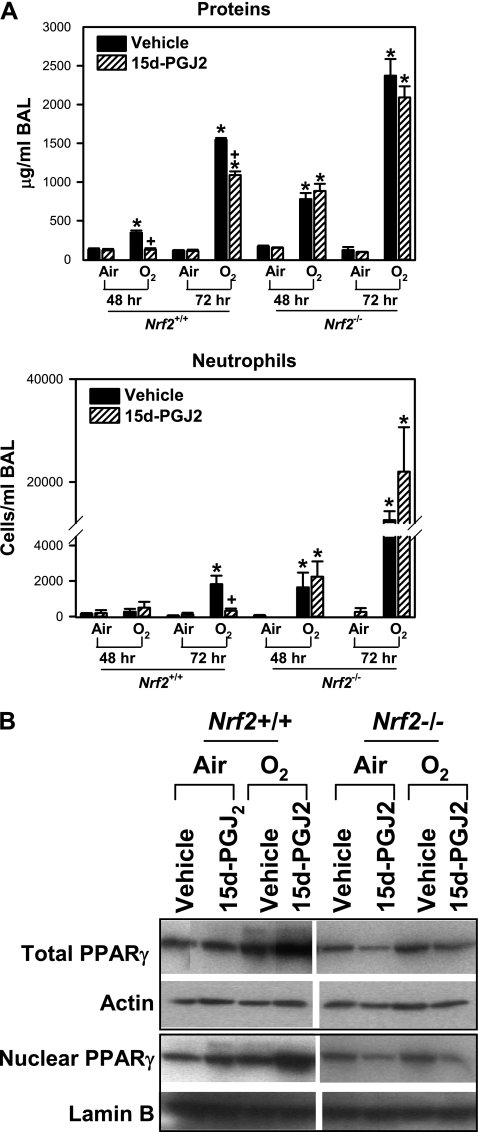
Rationale: The NF-E2 related factor 2 (Nrf2)-antioxidant response element (ARE) pathway is essential for protection against oxidative injury and inflammation including hyperoxia-induced acute lung injury. Microarray expression profiling revealed that lung peroxisome proliferator activated receptor gamma (PPARgamma) induction is suppressed in hyperoxia-susceptible Nrf2-deficient (Nrf2(-/-)) mice compared with wild-type (Nrf2(+/+)) mice. PPARgamma has pleiotropic beneficial effects including antiinflammation in multiple tissues.

Objectives: We tested the hypothesis that PPARgamma is an important determinant of pulmonary responsivity to hyperoxia regulated by Nrf2.

Methods: A computational bioinformatic method was applied to screen potential AREs in the Pparg promoter for Nrf2 binding. The functional role of a potential ARE was investigated by in vitro promoter analysis. A role for PPARgamma in hyperoxia-induced acute lung injury was determined by temporal silencing of PPARgamma via intranasal delivery of PPARgamma-specific interference RNA and by administration of a PPARgamma ligand 15-deoxy-Delta(12,14)-prostaglandin J(2) in mice.

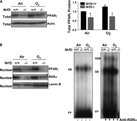
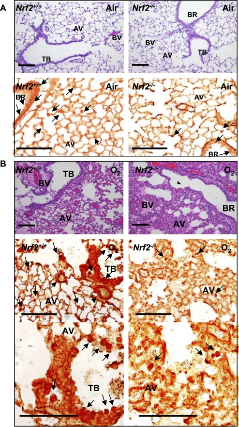
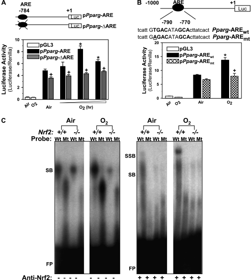
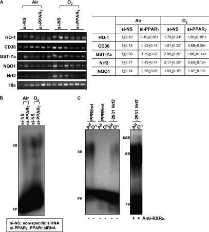
Measurements and main results: Deletion or site-directed mutagenesis of a potential ARE spanning -784/-764 sequence significantly attenuated hyperoxia-increased Pparg promoter activity in airway epithelial cells overexpressing Nrf2, indicating that the -784/-764 ARE is critical for Nrf2-regulated PPARgamma expression. Mice with decreased lung PPARgamma by specific interference RNA treatment had significantly augmented hyperoxia-induced pulmonary inflammation and injury. 15 Deoxy-Delta(12,14)-prostaglandin J(2) administration significantly reduced hyperoxia-induced lung inflammation and edema in Nrf2(+/+), but not in Nrf2(-/-) mice.

Conclusions: Results indicate for the first time that Nrf2-driven PPARgamma induction has an essential protective role in pulmonary oxidant injury. Our observations provide new insights into the therapeutic potential of PPARgamma in airway oxidative inflammatory disorders.


Figures

Figure 1.
Figure 1.
Pulmonary peroxisome proliferator activated receptor γ (PPARγ) level and activity was suppressed in Nrf2-deficient mice. (A) Aliquots of lung total protein were subjected for Western blotting to determine differential protein levels of pulmonary PPARγ in Nrf2+/+ and Nrf2−/− mice after exposure to air and O2 (48 h). Representative images are presented, and group mean ± SEM (n = 3/group) of total PPARγ level normalized to air-exposed Nrf2+/+ mice is depicted in a graph. * = significantly higher than genotype-matched air controls (P < 0.05). + = significantly lower than exposure-matched Nrf2+/+ mice (P < 0.05). (B) Aliquots of lung nuclear extracts isolated from pieces of left lung tissue (n = 3 mice/group) were subjected for Western blotting to determine differential nuclear translocation of pulmonary PPARγ and retinoid X receptor α protein in Nrf2+/+ and Nrf2−/− mice after exposure to air and O2 (48 h). Representative images are presented (n = 3/group). Differential nuclear protein–PPAR response element (PPRE) binding activity in the lungs of Nrf2+/+ and Nrf2−/− mice after exposure to air and O2 (48 h) was determined by gel shift/supershift analysis. Aliquots of nuclear protein were incubated with an end-labeled oligonucleotide probe containing PPRE consensus sequence. Total PPRE binding (shifted bands, left panel) and specific retinoid X receptor α–PPRE binding (super shifted bands, right panel) was determined by gel shift analysis. FP = free probes; RXRα = retinoid X receptor α; SB = shifted bands; SSB = super shifted bands. Representative images from multiple analysis (n = 3/group) are presented.
Figure 2.
Figure 2.
Pulmonary cellular localization of peroxisome proliferator activated receptor γ (PPARγ). Pulmonary histopathology demonstrated by hematoxylin and eosin staining and PPARγ localization determined by immunohistochemical staining of paraffin-embedded lung sections after 48 hour exposure to air (A) or O2 (B). Hyperoxia-induced protein edema in peribronchiolar and perivascular regions and alveolar air space, epithelial proliferation, and inflammatory cell infiltration shown in hematoxylin and eosin stained sections (A, B, upper panels) was markedly greater in Nrf2−/− mice relative to Nrf2+/+ mice. Cellular PPARγ localized by immunohistochemical staining (A, B, bottom panels) using an anti-PPARγ antibody indicated inflammatory cells and bronchiolar epithelial cells (arrows) as the primary sources of PPARγ in the hyperoxia-injured lung. PPARγ-positive cells were more predominantly enhanced in Nrf2+/+ mice relative to Nrf2−/− mice after hyperoxia. Representative light photomicrographs are shown (n = 3/group). Higher magnification photomicrographs displayed fewer occurrences of PPARγ-bearing cells in Nrf2−/− mice relative to Nrf2+/+ mice after O2. AV = alveoli; BR = bronchi or bronchiole; BV = blood vessel; TB = terminal bronchioles. Bars indicate 100 μm.
Figure 3.
Figure 3.
Functional assessment of a putative antioxidant response elements (ARE) on -784 region of Pparg promoter. (A) Transactivating activity of -784 ARE sequence was determined by promoter deletion analysis. Nrf2 overexpressing airway epithelial cells (BEAS-2B) were prepared by transfection with murine Nrf2 expression vector (pFlag-Nrf2). The cells were then transfected by Pparg promoter-luciferase reporter constructs with (pPparg-ARE) or without (pPparg-ΔARE) -784 ARE region, and exposed to either air or hyperoxia (–12 h). Mean ± SEM. presented (n = 6–9/group). * = significantly higher than air exposure under same transfection condition (P < 0.05). + = significantly lower than exposure-matched pPparg-ARE transfection (P < 0.05). (B) Transactivating activity of -784 ARE sequence was further determined by mutation analysis. Briefly, BEAS-2B cells overexpressing Nrf2 were transfected with a vector containing wild-type Pparg promoter bearing -784 ARE (pPparg-AREwt) or a vector containing the promoter with a mutation in -784 (pPparg-AREmt) incorporated by site-directed mutagenesis. The cells were then exposed to either air or hyperoxia (12 h). * = significantly higher than air exposure under the same transfection condition (P < 0.05). + = significantly lower than exposure-matched pPparg-AREwt (P < 0.05). (C) Gel shift analysis was performed on an aliquot of lung nuclear proteins from Nrf2+/+ and Nrf2−/− mice exposed to air or O2 (48 h) using γP32-end labeled wild-type (Wt; tcattGTGACataGCActtatcact) or mutated (Mt; tcattGGTACataGCActtatcact) -784 ARE-like probe. Hyperoxia increased Wt probe binding activity of nuclear proteins from Nrf2+/+, but not those from Nrf2−/− mice. Hyperoxia-induced increase of total DNA binding and specific Nrf2 binding (+ anti-Nrf2 antisera) was detected only on Wt probe by nuclear proteins from Nrf2+/+ mice. FP = free probes; SB = shifted bands; SSB = super shifted bands.
Figure 4.
Figure 4.
In vivo peroxisome proliferator activated receptor γ (PPARγ) specific interference RNA (siRNA) treatment inhibited pulmonary PPARγ expression and activity. (A) Aliquots of lung total and nuclear proteins were used for Western blotting to determine differential protein levels of pulmonary PPARγ in mice treated with nonspecific siRNA (si-NS) or PPARγ siRNA (si-PPARγ) followed by air and O2 exposure (72 h). Representative images from multiple analyses are presented, and group mean ± SEM (n = 3/group) of total and nuclear PPARγ normalized to air-exposed NS-treated mice are depicted in graphs. * = significantly higher than treatment-matched air controls (P < 0.05). + = significantly lower than exposure-matched NS-treated mice (P < 0.05). (B) Differential nuclear protein PPAR response element (PPRE) binding activity in the lungs of mice treated with si-NS or si-PPARγ after exposure to air and O2 (72 h). Aliquots of nuclear protein isolated from pieces of left lung tissue (n = 3 mice/group) were incubated with an end-labeled oligonucleotide probe containing PPRE consensus sequence. Total PPRE binding was determined by gel shift analysis. FP = free probes; SB = shifted bands of total bindings (PPRE motif-protein complex). Representative images from multiple analysis (n = 3) are presented.
Figure 5.
Figure 5.
In vivo peroxisome proliferator activated receptor γ (PPARγ) specific interference RNA (siRNA) treatment augmented pulmonary inflammation after hyperoxia. (A) Bronchopulmonary lavage (BAL) analysis determined the number of lung neutrophils, lymphocytes, and eosinophils in mice treated with nonspecific siRNA (si-NS) or PPARγ siRNA (si-PPARγ) followed by air and O2 exposure (72 h). Data are presented as group mean ± SEM (n = 3–6/group). (B) Transcription factor ELISA quantified specific DNA binding activity of nuclear p65 NF-κB. Group mean ± SEM (n = 3/group) normalized to air-exposed mice treated with si-NS are presented. (C) BAL levels of a proinflammatory cytokine IL-6 were determined by ELISA. Data presented as group mean ± SEM (n = 4–6/group). * = significantly higher than treatment-matched air controls (P < 0.05). + = significantly higher than O2-exposed si-NS-treated mice (P < 0.05).
Figure 6.
Figure 6.
In vivo peroxisome proliferator activated receptor γ (PPARγ) specific interference RNA (siRNA) treatment suppressed pulmonary apoptosis markers after hyperoxia. (A) Terminal deoxynucleotidyl transferase-mediated dUTP nick-end labeling (TUNEL) assay determined cells undergoing apoptosis in lung tissue sections after air or hyperoxia exposure (72 h). Hyperoxia caused cellular apoptosis predominantly in inflammatory, endothelial, and epithelial cells in all mice. Intensity and abundance of TUNEL-positive cells increased by hyperoxia were greater in mice treated with nonspecific siRNA (si-NS) compared with mice treated with PPARγ-specific siRNA (si-PPARγ). Higher magnification photomicrographs displayed differentially less abundant apoptotic macrophages, epithelial cells, and endothelial cells in hyperoxia-exposed lungs treated with si-PPARγ than in lungs treated with si-NS. AV = alveoli; BR = bronchi or bronchiole; BV = blood vessel; TB = terminal bronchioles. Arrows indicate TUNEL-stained nuclei. Bars indicate 100 μm. Representative light photomicrographs are shown (n = 3/group). (B) O2-enhanced lung levels of PPARγ-mediated apoptosis signaling proteins, PTEN, caspase-8, and bax determined by Western blot analysis in aliquots of total lung proteins were greater in mice treated with si-NS than in mice treated with si-PPARγ (72 h). Representative band images are shown (n = 3/group).
Figure 7.
Figure 7.
In vivo peroxisome proliferator activated receptor γ (PPARγ) specific interference RNA (siRNA) treatment suppressed cytoprotective gene expression and antioxidant response elements (ARE) binding activity after hyperoxia. (A) Message levels of cytoprotective genes HO-1, CD36, GST-Ya, and NQO1 and Nrf2 were detected by semiquantitative reverse transcriptase–polymerase chain reaction using total lung RNA isolated from mice treated with nonspecific siRNA (si-NS) or PPARγ-specific siRNA (si-PPARγ) after air and O2 exposure (72 h). cDNA band images for each gene and quantified relative intensities to air-exposed NS-treated mice of digitized cDNA bands normalized to the intensity of each 18s band are shown. Data are presented as group mean ± SEM (n = 3/group). * = significantly higher than treatment-matched air controls (P < 0.05). + = significantly lower than exposure-matched si-NS-treated mice (P < 0.05). (B) Differential nuclear protein-ARE binding activity in the lungs of mice treated with si-NS or si-PPARγ after exposure to air and O2 (72 h). Aliquots of nuclear protein isolated from pooled pieces of left lung tissues (n = 3 mice/group) were incubated with an end-labeled oligonucleotide probe containing ARE consensus sequence. Total ARE binding was determined by gel shift analysis. FP = free ARE probes; SB = shifted bands of total bindings (ARE motif-protein complex). Representative images from multiple analysis (n = 2) are presented. (C) Increased total nuclear protein binding activity (SB) and specific PPARγ-retinoic acid X receptor (RXRa) binding activity (SSB) on an Nrf2 PPAR response element (PPRE)–like sequence in the lungs of mice after exposure to O2 (72 h). Aliquots of nuclear protein isolated from pooled pieces of left lung tissue exposed to either air or O2 (n = 3 mice/group) were incubated with an end-labeled oligonucleotide probe containing -2931 Nrf2 PPRE-like sequence (-2931 Nrf2). PPRE consensus sequence (PPREwt) was used as a positive control for band shift, and mutant PPRE (PPREmt) as a no binding control. FP = free DNA probes; SB = shifted bands of total bindings (PPRE motif-protein complex); SSB = super shifted bands of specific RXRα bindings (PPRE motif-protein–anti-RXRα antibody complex). Representative images from multiple analysis (n = 2) are presented.
Figure 8.
Figure 8.
Treatment with 15-deoxy-Δ12,14-prostaglandin J2 (15d-PGJ2) enhanced lung peroxisome proliferator activated receptor γ (PPARγ) expression and suppressed pulmonary injury caused by hyperoxia in Nrf2+/+ mice. (A) Bronchopulmonary lavage (BAL) analysis determined the concentration of total proteins and the number of lung neutrophils in Nrf2+/+ and Nrf2−/− mice pretreated with either vehicle or 15d-PGJ2 followed by air and O2 exposure (48 and 72 h). Data are presented as group mean ± SEM (n = 3–7/group). * = significantly higher than genotype- and treatment-matched air controls (P < 0.05). + = significantly lower than genotype- and exposure-matched vehicle-treated mice (P < 0.05). (B) Aliquots of lung total and nuclear proteins were subjected for Western blotting to determine PPARγ levels in Nrf2+/+ and Nrf2−/− mice treated with vehicle or 15d-PGJ2 after air or O2 exposure (72 h). Representative images from multiple analysis (n = 3 for total proteins, n = 2 for nuclear proteins) are presented.

Comment in

Similar articles

Cited by

References

    1. Bowler RP, Barnes PJ, Crapo JD. The role of oxidative stress in chronic obstructive pulmonary disease. COPD 2004;1:255–277. - PubMed
    1. Chow CW, Herrera Abreu MT, Suzuki T, Downey GP. Oxidative stress and acute lung injury. Am J Respir Cell Mol Biol 2003;29:427–431. - PubMed
    1. Paz-Elizur T, Krupsky M, Elinger D, Schechtman E, Livneh Z. Repair of the oxidative DNA damage 8-oxoguanine as a biomarker for lung cancer risk. Cancer Biomark 2005;1:201–205. - PubMed
    1. Ware LB, Matthay MA. The acute respiratory distress syndrome. N Engl J Med 2000;342:1334–1349. - PubMed
    1. Itoh K, Chiba T, Takahashi S, Ishii T, Igarashi K, Katoh Y, Oyake T, Hayashi N, Satoh K, Hatayama I, et al. An Nrf2/small MAF heterodimer mediates the induction of phase II detoxifying enzyme genes through antioxidant response elements. Biochem Biophys Res Commun 1997;236:313–322. - PubMed

Publication types