. 2010 Jun;9(6):1324-38.
doi: 10.1074/mcp.M000063-MCP201. Epub 2010 Mar 11.

Proteomics analysis of bladder cancer exosomes

Affiliations

Proteomics analysis of bladder cancer exosomes

Joanne L Welton et al. Mol Cell Proteomics. 2010 Jun.

Abstract

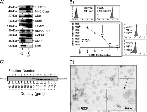
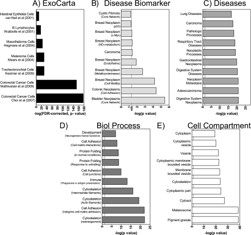
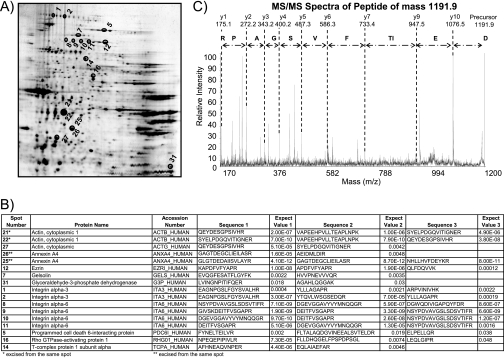
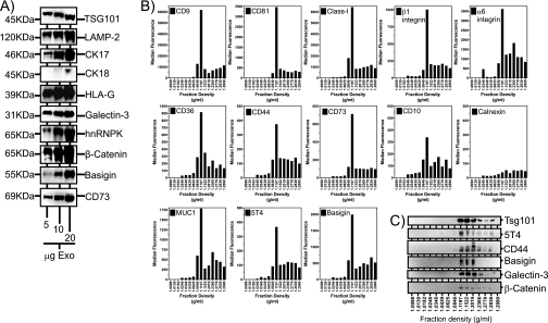
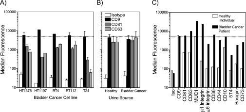
Exosomes are nanometer-sized vesicles, secreted by various cell types, present in biological fluids that are particularly rich in membrane proteins. Ex vivo analysis of exosomes may provide biomarker discovery platforms and form non-invasive tools for disease diagnosis and monitoring. These vesicles have never before been studied in the context of bladder cancer, a major malignancy of the urological tract. We present the first proteomics analysis of bladder cancer cell exosomes. Using ultracentrifugation on a sucrose cushion, exosomes were highly purified from cultured HT1376 bladder cancer cells and verified as low in contaminants by Western blotting and flow cytometry of exosome-coated beads. Solubilization in a buffer containing SDS and DTT was essential for achieving proteomics analysis using an LC-MALDI-TOF/TOF MS approach. We report 353 high quality identifications with 72 proteins not previously identified by other human exosome proteomics studies. Overrepresentation analysis to compare this data set with previous exosome proteomics studies (using the ExoCarta database) revealed that the proteome was consistent with that of various exosomes with particular overlap with exosomes of carcinoma origin. Interrogating the Gene Ontology database highlighted a strong association of this proteome with carcinoma of bladder and other sites. The data also highlighted how homology among human leukocyte antigen haplotypes may confound MASCOT designation of major histocompatability complex Class I nomenclature, requiring data from PCR-based human leukocyte antigen haplotyping to clarify anomalous identifications. Validation of 18 MS protein identifications (including basigin, galectin-3, trophoblast glycoprotein (5T4), and others) was performed by a combination of Western blotting, flotation on linear sucrose gradients, and flow cytometry, confirming their exosomal expression. Some were confirmed positive on urinary exosomes from a bladder cancer patient. In summary, the exosome proteomics data set presented is of unrivaled quality. The data will aid in the development of urine exosome-based clinical tools for monitoring disease and will inform follow-up studies into varied aspects of exosome manufacture and function.


Figures

Fig. 1.
Fig. 1.
Characterization of HT1376-derived exosomes using Western blotting, flow cytometry, and electron microscopy. Cell (CL) and exosome (Exo) lysates (5 μg/well) were compared by Western blotting using a range of antibodies as indicated. This demonstrated relative enrichment of several proteins in exosomes. Some markers, such as gp96, were absent from exosomes, indicating negligible contamination of the preparations by cellular debris (this is representative of three experiments) (A). Exosomes coupled to latex beads were analyzed by flow cytometry, and this revealed positive expression of tetraspanin molecules on the exosome surface. Median fluorescence intensity values (MFI) are shown (representative of >5 experiments) (B). Intentional contamination of purified exosomes with increasing amounts of FBS prior to coupling to latex beads reveals a decrease in signal intensity for CD9 (mean ± S.E.; n = 6; **, p < 0.001, one-way analysis of variance with Tukey's post test) (B, line graph). Material pelleted at 70,000 × g from cell-conditioned medium was overlaid on a linear sucrose gradient (0.2–2.02 m) and ultracentrifuged for 18 h at 210,000 × g. Collected fractions were analyzed by refractometry to ascertain fraction density and thereafter by Western blot using antibodies to TSG101, which is an exosome marker. TSG101 floats at typical exosome densities of between 1.1 and 1.2 g/ml (representative of four experiments) (C). Transmission electron micrograph of a typical exosome preparation reveals heterogeneous vesicles between 30 and 100 nm in diameter (D). CK, cytokeratin.
Fig. 2.
Fig. 2.
Summary of overrepresentation analysis of nano-LC/MS-derived protein identifications against gene sets from ExoCarta and GeneGO. To facilitate comparison with ExoCarta gene sets, our protein list was first converted to an EntrezGene-identified gene list before undertaking ORA using the hypergeometric distribution. Results were filtered to include comparisons with MS-based studies only and with those reporting 10 or more matching genes, yielding seven studies (, –43). This demonstrates how well our MS data compare with exosome protein profiles from specified cell types, displayed as the −log(p value) corrected for false detection rate (A). ORA analysis using MetaCore utilized the Swiss-Prot IDs for the identified protein list. For clarity, we report the top 10 overrepresented genes contained within each of the following group headings: disease biomarker (B), diseases (C), biological (Biol) process (D), and cellular compartment (E). The dotted line indicates p = 0.05; hence, columns to the left of this are not statistically significant (ns).
Fig. 3.
Fig. 3.
Analysis of HT1376-derived exosomes using 2DE and MS. Protein extracts from HT1376 exosomes were resolved by 2DE on a pH 3–10 non-linear gradient. Proteins were visualized by silver staining (A). Thirty-two spots were randomly chosen, gel plugs were excised, and peptides were recovered following trypsin digestion. Of these, successful identifications were obtained for 17 spots (annotated in A), and the details of the MS identifications are listed (B). A representative MS/MS analysis from the data set is shown in C; the peptide is from integrin α6 (spot 10). The peptide has a precursor mass of 1191.9 and is annotated to show the derived peptide sequence.
Fig. 4.
Fig. 4.
Validation of some MS-identified proteins by Western blot and flow cytometric analysis. HT1376 exosomes (5–20 μg/well), purified by the standard sucrose cushion method, were analyzed by Western blot for expression of a range of MS identified proteins as indicated (A). The 70,000 × g pellet, obtained from HT1376 cell-conditioned medium, was subjected to fractionation by centrifugation on a linear sucrose gradient (0.2–2.5 m). Fifteen total fractions were collected, and the density was measured by refractometry. Thereafter, one-third of each fraction was coupled to latex beads followed by flow cytometric analysis for exosomal surface expression as indicated (B). In parallel, the remaining two-thirds of each fraction was subjected to Western blotting for proteins as indicated (C). The data reveal proteins floating at a recognized exosomal density range (1.12–1.2 g/ml). (The data are representative of two experiments.) CK, cytokeratin.
Fig. 5.
Fig. 5.
Exosomes isolated from urine specimens. The sucrose cushion method, used in this study for HT1376 exosomes, was also tested on a small panel of other bladder cancer cell lines, and the quality of preparations was assessed using the latex bead assay (A). The graph depicts median fluorescence values from the flow cytometric histogram peak (mean ± S.D. of n preparations where n = 5 for HT1376, n = 1 for HT1197, n = 2 for RT4, n = 2 for RT112, and n = 2 for T24) with beads stained for CD9, CD81, or CD63 as indicated. The dotted line indicates our arbitrary cutoff value (of 5000 units) where CD9 fluorescence above this value was indicative of a preparation of acceptable quality. Using identical methods, exosomes were prepared from urine specimens collected from healthy individuals (n = 4) or from bladder cancer patients (n = 3) (mean ± S.D.), and exosomes were analyzed as above (B). Exosome preparations from one healthy individual and one bladder cancer patient, which exceeded the quality threshold, were analyzed further for surface expression of some MS-identified proteins as indicated (C). Iso, isotype.

References

    1. Caby M. P., Lankar D., Vincendeau-Scherrer C., Raposo G., Bonnerot C. (2005) Exosomal-like vesicles are present in human blood plasma. Int. Immunol 17, 879–887 - PubMed
    1. Pisitkun T., Shen R. F., Knepper M. A. (2004) Identification and proteomic profiling of exosomes in human urine. Proc. Natl. Acad. Sci. U.S.A 101, 13368–13373 - PMC - PubMed
    1. Admyre C., Johansson S. M., Qazi K. R., Filén J. J., Lahesmaa R., Norman M., Neve E. P., Scheynius A., Gabrielsson S. (2007) Exosomes with immune modulatory features are present in human breast milk. J. Immunol 179, 1969–1978 - PubMed
    1. Gatti J. L., Métayer S., Belghazi M., Dacheux F., Dacheux J. L. (2005) Identification, proteomic profiling, and origin of ram epididymal fluid exosome-like vesicles. Biol. Reprod 72, 1452–1465 - PubMed
    1. Bard M. P., Hegmans J. P., Hemmes A., Luider T. M., Willemsen R., Severijnen L. A., van Meerbeeck J. P., Burgers S. A., Hoogsteden H. C., Lambrecht B. N. (2004) Proteomic analysis of exosomes isolated from human malignant pleural effusions. Am. J. Respir. Cell Mol. Biol 31, 114–121 - PubMed

Publication types

MeSH terms

Substances