

Alpha-synuclein deficient mice are resistant to toxin-induced multiple system atrophy
- PMID: 20224454
- PMCID: PMC3049936
- DOI: 10.1097/WNR.0b013e328338ba6b
Alpha-synuclein deficient mice are resistant to toxin-induced multiple system atrophy
Abstract
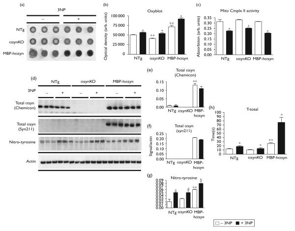
Multiple systems atrophy (MSA) is a neurodegenerative disorder characterized by oligodendrocytic accumulations of alpha-synuclein (alphasyn). Oxidative stress is a key mechanism proposed to underlie MSA pathology. To address the role of alphasyn modifications, over and above general oxidative modifications, this study examined the effects of 3-nitropropionic acid (3NP) administration, a technique used to model MSA, in knock-out mice lacking alphasyn (alphasynKO). Although susceptible to 3NP-induced oxidative stress, alphasynKO mice display reduced neuronal loss and dendritic pathology. The alphasynKO mice are resistant to 3NP-induced motor deficits and display attenuated loss of tyrosine hydroxylase and dopamine transporter striatal immunoreactivity. The results suggest that deficits in MSA are not due to general oxidative protein modification but in addition may be related to specific alphasyn modifications.
Figures


Similar articles
-
Mitochondrial inhibitor 3-nitroproprionic acid enhances oxidative modification of alpha-synuclein in a transgenic mouse model of multiple system atrophy.J Neurosci Res. 2009 Sep;87(12):2728-39. doi: 10.1002/jnr.22089. J Neurosci Res. 2009. PMID: 19405128 Free PMC article.
-
Failure of Neuroprotection Despite Microglial Suppression by Delayed-Start Myeloperoxidase Inhibition in a Model of Advanced Multiple System Atrophy: Clinical Implications.Neurotox Res. 2015 Oct;28(3):185-94. doi: 10.1007/s12640-015-9547-7. Epub 2015 Jul 21. Neurotox Res. 2015. PMID: 26194617 Free PMC article.
-
Neurodegeneration in a transgenic mouse model of multiple system atrophy is associated with altered expression of oligodendroglial-derived neurotrophic factors.J Neurosci. 2010 May 5;30(18):6236-46. doi: 10.1523/JNEUROSCI.0567-10.2010. J Neurosci. 2010. PMID: 20445049 Free PMC article.
-
Is Multiple System Atrophy a Prion-like Disorder?Int J Mol Sci. 2021 Sep 18;22(18):10093. doi: 10.3390/ijms221810093. Int J Mol Sci. 2021. PMID: 34576255 Free PMC article. Review.
-
Animal models of multiple system atrophy.Neuroscience. 2012 Jun 1;211:77-82. doi: 10.1016/j.neuroscience.2011.09.044. Epub 2011 Sep 25. Neuroscience. 2012. PMID: 21963351 Review.
Cited by
-
A novel triple repeat mutant tau transgenic model that mimics aspects of pick's disease and fronto-temporal tauopathies.PLoS One. 2015 Mar 24;10(3):e0121570. doi: 10.1371/journal.pone.0121570. eCollection 2015. PLoS One. 2015. Retraction in: PLoS One. 2025 May 28;20(5):e0325329. doi: 10.1371/journal.pone.0325329. PMID: 25803611 Free PMC article. Retracted.
-
Multiple system atrophy: a clinical and neuropathological perspective.Trends Neurosci. 2011 Nov;34(11):581-90. doi: 10.1016/j.tins.2011.08.003. Epub 2011 Sep 29. Trends Neurosci. 2011. PMID: 21962754 Free PMC article. Review.
-
Multiple system atrophy: current and future approaches to management.Ther Adv Neurol Disord. 2010 Jul;3(4):249-63. doi: 10.1177/1756285610375328. Ther Adv Neurol Disord. 2010. PMID: 21179616 Free PMC article.
-
Multiple system atrophy: pathogenic mechanisms and biomarkers.J Neural Transm (Vienna). 2016 Jun;123(6):555-72. doi: 10.1007/s00702-016-1545-2. Epub 2016 Apr 20. J Neural Transm (Vienna). 2016. PMID: 27098666 Review.
-
LIMP-2 expression is critical for β-glucocerebrosidase activity and α-synuclein clearance.Proc Natl Acad Sci U S A. 2014 Oct 28;111(43):15573-8. doi: 10.1073/pnas.1405700111. Epub 2014 Oct 14. Proc Natl Acad Sci U S A. 2014. PMID: 25316793 Free PMC article.
References
-
- Hanna PA, Jankovic J, Kirkpatrick JB. Multiple system atrophy: the putative causative role of environmental toxins. Arch Neurol. 1999;56:90–94. - PubMed
-
- Nee LE, Gomez MR, Dambrosia J, Bale S, Eldridge R, Polinsky RJ. Environmental-occupational risk factors and familial associations in multiple system atrophy: a preliminary investigation. Clin Auton Res. 1991;1:9–13. - PubMed
-
- Vanacore N, Bonifati V, Fabbrini G, Colosimo C, De Michele G, Marconi R, et al. Case-control study of multiple system atrophy. Mov Disord. 2005;20:158–163. - PubMed
-
- Norris EH, Giasson BI, Ischiropoulos H, Lee VM. Effects of oxidative and nitrative challenges on alpha-synuclein fibrillogenesis involve distinct mechanisms of protein modifications. J Biol Chem. 2003;278:27230–27240. - PubMed
Publication types
MeSH terms
Substances
Grants and funding
LinkOut - more resources
Full Text Sources
Other Literature Sources
