. 2010 Apr 21;29(8):1434-45.
doi: 10.1038/emboj.2010.27. Epub 2010 Mar 11.

A cooperative activation loop among SWI/SNF, gamma-H2AX and H3 acetylation for DNA double-strand break repair

Affiliations

A cooperative activation loop among SWI/SNF, gamma-H2AX and H3 acetylation for DNA double-strand break repair

Han-Sae Lee et al. EMBO J. .

Abstract

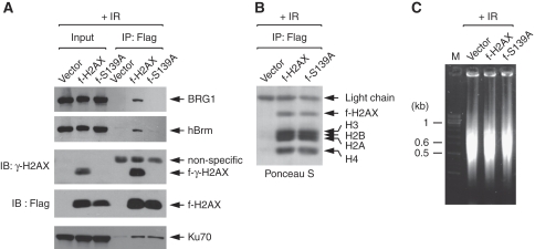
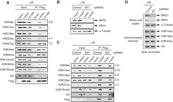
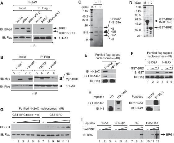
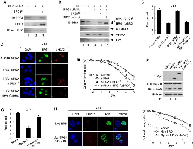
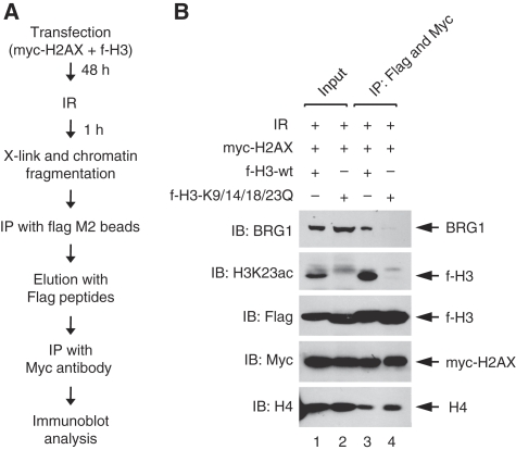
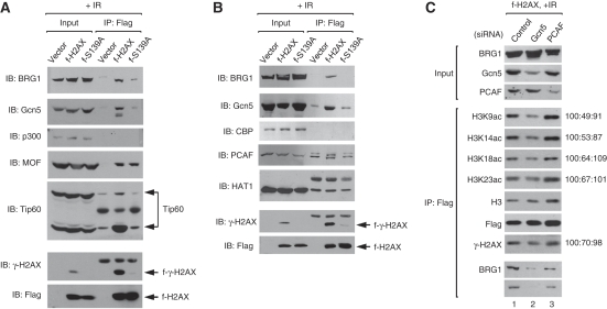
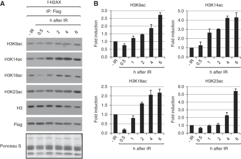
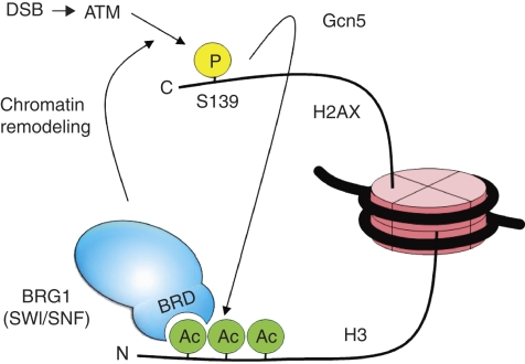
Although recent studies highlight the importance of histone modifications and ATP-dependent chromatin remodelling in DNA double-strand break (DSB) repair, how these mechanisms cooperate has remained largely unexplored. Here, we show that the SWI/SNF chromatin remodelling complex, earlier known to facilitate the phosphorylation of histone H2AX at Ser-139 (S139ph) after DNA damage, binds to gamma-H2AX (the phosphorylated form of H2AX)-containing nucleosomes in S139ph-dependent manner. Unexpectedly, BRG1, the catalytic subunit of SWI/SNF, binds to gamma-H2AX nucleosomes by interacting with acetylated H3, not with S139ph itself, through its bromodomain. Blocking the BRG1 interaction with gamma-H2AX nucleosomes either by deletion or overexpression of the BRG1 bromodomain leads to defect of S139ph and DSB repair. H3 acetylation is required for the binding of BRG1 to gamma-H2AX nucleosomes. S139ph stimulates the H3 acetylation on gamma-H2AX nucleosomes, and the histone acetyltransferase Gcn5 is responsible for this novel crosstalk. The H3 acetylation on gamma-H2AX nucleosomes is induced by DNA damage. These results collectively suggest that SWI/SNF, gamma-H2AX and H3 acetylation cooperatively act in a feedback activation loop to facilitate DSB repair.


Conflict of interest statement

The authors declare that they have no conflict of interest.

Figures

Figure 1
Figure 1
SWI/SNF binds to γ-H2AX nucleosomes in S139ph-dependent manner. (A) Flag-tagged nucleosomes were immunoprecipitated from the cells stably expressing either f-H2AX or f-S139A at 1 h after 10-Gy IR, and analysed for flag-tagged histones, γ-H2AX and associating proteins by immunoblot with specific antibodies. As control, the cells containing empty vector were subjected in parallel to the nucleosome immunoprecipitation. (B) Ponceau S stain of immunoblot shows that immunoprecipitated f-H2AX nucleosomes are intact. (C) DNA of fragmented chromatin was analysed along with a size marker (M) on agarose gel.
Figure 2
Figure 2
S139ph is required for the acetylation of H3 on γ-H2AX nucleosomes. (A) Flag-tagged nucleosomes were immunoprecipitated from the cells containing empty vector, or expressing either f-H2AX or f-S139A at 1 h after 10-Gy IR, and analysed for the acetylations and methylations of H3 by immunoblot with specific antibodies. The bands of acetyl-H3 on precipitated f-H2AX and f-S139ph nucleosomes were quantitated by densitometer, and after normalization to H3 bands, the fold reduction of H3 acetylation was calculated and shown at the right side of the corresponding gel. For the gel with star mark, the fold reduction was not calculable as the band intensity of f-S139A lane is lower than background. The nomenclature of histone modifications used in this paper was followed by Turner (2005); ac, acetylation; me, monomethylation; me2, dimethylation; ph, phosphorylation. (B) Vector and f-H2AX cells were transfected with either the siRNAs specific for BRG1 or hBrm (B/h), or non-specific control siRNA. Cells were collected at 1 h after 10-Gy IR for the analysis of BRG1 and hBrm knockdown by immunoblot with specific antibodies; α-tubulin was also analysed for internal control. (C) Flag-tagged nucleosomes were immunoprecipitated from the aliquots of the cells prepared in (B) were analysed for the indicated modifications of H3 by immunoblot using specific antibodies. The fold reduction of H3 acetylation on precipitated f-H2AX nucleosomes by SWI/SNF knockdown was calculated as per in (A) and shown at the right side of the corresponding gel. (D) Cells were transfected with either control or BRG1/hBrm-specific siRNAs, and at 1 h after 10-Gy IR, whole cell lysates were prepared for the analysis of BRG1 and hBrm expression, and acid-extracted histones for the analysis of H3 acetylation as indicated.
Figure 3
Figure 3
BRG1 binds to γ-H2AX nucleosomes by interacting with acetylated H3 through its bromodomain. (A) f-H2AX cells were transfected with empty vector, or the expression vectors for the full-length BRG1 or the BRD-deleted BRG1 (BRG1ΔBRD). Flag-tagged nucleosomes were immunoprecipitated at 1 h after 10-Gy IR and analysed for associating proteins by immunoblot. (B) f-H2AX and f-S139A cells were transfected with empty vector (V) or the expression vector for myc-tagged BRG1 BRD (Myc-BRD, b). Flag-tagged nucleosomes were immunoprecipitated at 1 h after 10-Gy IR and analysed for associating proteins by immunoblot. Non-specific (NS) bands are also indicated. (C) f-H2AX (lane 1) and f-S139A (lane 2) cells were exposed to 10-Gy IR, and after 1 h, cells were collected and subjected to the affinity purification of flag-tagged nucleosomes. Coomassie stain gel of the purified nucleosomes is shown. (D) The GST proteins containing the 588–748 aa of BRG1 (lane 1) or the BRG1 BRD (lane 2) were expressed and purified from Escherichia coli, and analysed on an SDS gel with coomassie stain. (E) The purified flag-tagged nucleosomes shown in (C) were analysed for γ-H2AX and H3K14ac by immunoblot. (F) Affinity-purified f-S139A and f-H2AX nucleosomes were incubated with buffer only (lanes 1 and 4) or GST-BRD at increasing molar ratios of 1:2 (lanes 2 and 5) or 1:8 (lanes 3 and 6). Flag-tagged nucleosomes were immunoprecipitated and analysed for associating proteins by immunoblot. (G) Affinity-purified f-H2AX nucleosomes were incubated with buffer only (lanes 1 and 7) or indicated GST proteins at increasing molar ratios of 1:1 (lanes 2 and 8), 1:2 (lanes 3 and 9), 1:4 (lanes 4 and 10), 1:8 (lanes 5 and 11) or 1:16 (lanes 6 and 12). The nucleosomes were immunoprecipitated and analysed for associating proteins by immunoblot. (H) Verification of synthetic peptides by immunoblot analysis. Unmodified and K14-acetylated H3 peptides (left panel), and unmodified and S139-phosphorylated H2AX peptides (right panel) were run on 18% SDS gel and subjected to immunoblot with specific antibodies as indicated. (I) Indicated biotinylated peptides (5 μg/ml) were incubated with buffer only (lanes 1, 4, 7 and 10) or purified SWI/SNF complexes at the concentrations of 0.2 μg/ml (lanes 2, 5, 8 and 11) or 0.8 μg/ml (lanes 3, 6, 9 and 12). Peptide-protein complexes were pull downed by streptavidin-coated beads and the bead-bound proteins were analysed by immunoblot.
Figure 4
Figure 4
BRG1 binding to γ-H2AX nucleosomes is important for S139ph and DSB repair. (A) Cells were cotransfected with non-specific (lane 1) or BRG1-specific siRNAs (lanes 2 and 3), plus either empty vectors (lanes 1 and 2) or the expression vectors for HA-tagged siRNA-resistant BRG1 (BRG1R) (lane 3). Whole cell lysates were analysed for the expression of BRG1 and BRG1R by immunoblot. (B) Cells were cotransfected with non-specific (lanes 1 and 2) or BRG1-specific siRNAs (lanes 3–5), plus either empty vectors (lanes 1–3) or the expression vectors for BRG1R (lane 4) or BRD-deleted BRG1R (BRG1RΔBRG1, lane 5). At 1 h after untreated (lane 1) or 10-Gy IR (lanes 2–5), cells were collected to prepare whole cell lysates and acid-extracted histones for immunoblots with anti-BRG1 or anti-γ-H2AX antibodies, respectively; α-tubulin and H2A were also analysed for loading control. (C) Cells were transfected as described in lanes 2–5 of (B) and exposed to 2-Gy IR. After 1 h, cells were fixed and dually stained with anti-BRG1 or anti-γ-H2AX antibodies before confocal images were captured. Average number of γ-H2AX foci per cell was depicted as graph by counting at least 50 cells. The error bar indicates mean±s.d. of three independent experiments. (D) Representative confocal images from the experiments in (C) are shown. (E) Cells transfected as per in (D) were untreated (0 Gy) or exposed to 1–5 Gy IR before the viability was determined by colony formation assays with triplicates per sample. The graph shows average number of colonies with mean±s.d. of four independent experiments. (F) Cells were transfected with empty or Myc-BRD expression vectors. At 1 h after 10-Gy IR, cells were collected to prepare whole cell lysates and acid-extracted histones for immunoblots with anti-Myc or anti-γ-H2AX antibodies, respectively; α-tubulin and H2A were also analysed for loading control. (G) Cells transfected with empty, Myc-BRD or Myc-BRG1(588–748) vectors were irradiated by 2 Gy, and after 1 h, cells were fixed for dual staining with the antibodies against Myc or γ-H2AX. The Myc-BRG1(588–748) vector expresses the sequences of 588–748 amino acids of BRG1, outside the BRD, and was used as a control. Average number of γ-H2AX foci per cell was depicted as graph by counting at least 50 each of untransfected and transfected cells. The error bar indicates mean±s.d. of three independent experiments. (H) Representative confocal images from the experiments in (G) are shown. (I) Cells were transfected with indicated vectors, and after irradiation, cells were subjected to colony formation assays as described in (E). The graph shows average number of colonies with mean±s.d. of three independent experiments.
Figure 5
Figure 5
H3 acetylation is required for the binding of BRG1 to γ-H2AX nucleosomes. (A) The experimental procedure used in (B) is represented as a schematic flow chart. See Materials and methods for details. (B) 293T cells were transfected with myc-H2AX plus either f-H3-wt (lanes 1 and 3) or f-H3-K9/14/18/23Q vectors (lanes 2 and 4), and irradiated by 10 Gy 1 h before harvest. f-H2AX nucleosomes were immunoprecipitated using anti-Flag M2 beads and eluted with Flag peptides, and the Flag-eluted nucleosomes were then subjected to the second immunoprecipitation by Myc antibody followed by immunoblot analysis as indicated. The K to Q mutations for the four acetylation sites were verified by DNA sequencing (Materials and methods) as well as by immunoblot analysis with specific antibodies (here and data not shown). H4 was also analysed to monitor the integrity of the precipitated nucleosomes.
Figure 6
Figure 6
Gcn5 is responsible for the γ-H2AX-mediated H3 acetylation. (A, B) To search for the HATs that bind to γ-H2AX nucleosomes in the S139ph-specific manner, all the HATs earlier shown to be implicated in DNA repair were examined by the similar experiments as described in Figure 1. Several independent sets of experiments were performed to test some subgroups of HATs in each of which the specific interaction between BRG1 and γ-H2AX nucleosomes was always monitored to control the chromatin IP experiments. The results of two representative experimental sets are shown. The specific interaction between Gcn5 and γ-H2AX nucleosomes was also included in the experimental set shown in (B). (C) Gcn5 knockdown reduces the acetylation of H3 on γ-H2AX nucleosomes. f-H2AX cells were transfected with non-specific (lane 1), Gcn5- (lane 2) or PCAF-specific siRNAs (lane 3), and at 1 h after 10-Gy IR, cells were fixed and sonicated for the preparation of fragmented chromatin lysate (Input). f-H2AX nucleosomes were immunoprecipitated and divided into two to analyse the levels of H3 acetylation and the binding of BRG1 and Gcn5 to f-H2AX nucleosomes in separate gels as indicated. The levels of γ-H2AX on f-H2AX nucleosomes was also analysed using the same blot as used for the analysis of H3 acetylation. The input lysate was analysed for siRNA knockdown of Gcn5 and PCAF as well as for the levels of BRG1. The relative band intensity of acetyl-H3 and γ-H2AX after normalization to H3 and f-H2AX bands, respectively, is shown next to the corresponding gel.
Figure 7
Figure 7
IR induces H3 acetylation on γ-H2AX nucleosomes. (A) f-H2AX cells were left untreated (−IR) or irradiated by 10 Gy, and the irradiated cells were harvested after various times. f-H2AX nucleosomes were immunoprecipitated from those cells and subjected to immunoblot analysis using specific antibodies. The immunoblot membrane stained with Ponceau S before incubation with antibodies is shown below the gel. (B) The quantitation data for the gel in (A) is shown. The bands of acetyl-H3 were quantitated by densitometer, and after normalization to H3, the fold induction of H3 acetylation was calculated and shown next to the corresponding gel. The error bar indicates mean±s.d. of three independent experiments.
Figure 8
Figure 8
A model for the cooperative action of SWI/SNF, S139ph and H3 acetylation during DSB repair. For clarity, only BRG1 subunit is shown for the SWI/SNF complex. See text for details.

Similar articles

Cited by

References

    1. Agalioti T, Chen G, Thanos D (2002) Deciphering the transcriptional histone acetylation code for a human gene. Cell 111: 381–392 - PubMed
    1. Allard S, Masson JY, Cote J (2004) Chromatin remodeling and the maintenance of genome integrity. Biochim Biophys Acta 1677: 158–164 - PubMed
    1. Allis CD, Berger SL, Cote J, Dent S, Jenuwien T, Kouzarides T, Pillus L, Reinberg D, Shi Y, Shiekhattar R, Shilatifard A, Workman J, Zhang Y (2007) New nomenclature for chromatin-modifying enzymes. Cell 131: 633–636 - PubMed
    1. Bao Y, Shen X (2007) Chromatin remodeling in DNA double-strand break repair. Curr Opin Genet Dev 17: 126–131 - PubMed
    1. Bird AW, Yu DY, Pray-Grant MG, Qiu Q, Harmon KE, Megee PC, Grant PA, Smith MM, Christman MF (2002) Acetylation of histone H4 by Esa1 is required for DNA double-strand break repair. Nature 419: 411–415 - PubMed

Publication types