

Occlusion of Internal Carotid Artery in Kimura's Disease
- PMID: 20224649
- PMCID: PMC2833309
- DOI: 10.1155/2010/407538
Occlusion of Internal Carotid Artery in Kimura's Disease
Abstract
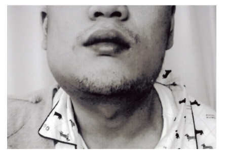
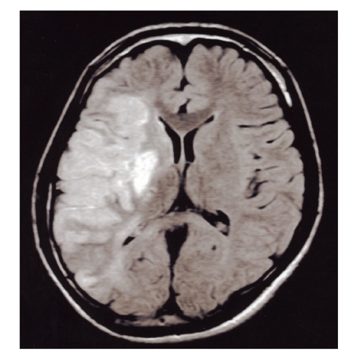
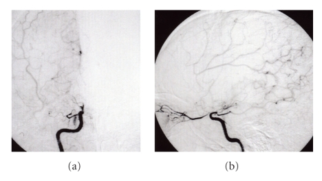
We describe a unique case of Kimura's disease in which cerebral infarction was caused by occlusion of the right internal carotid artery. A 25-year-old man with Kimura's disease was admitted to our hospital because of left hemiparesis. Computed tomography and magnetic resonance imaging of the head showed infarction in the right frontal and temporal lobes. Cerebral angiography demonstrated right internal carotid artery occlusion affecting the C1 segment, with moyamoya-like collateral vessels arising from the right opthalamic artery. Kimura's disease is a chronic disease characterized by the clinical triad of slowly enlarging subcutaneous masses with lymphoid hyperplasia in the head and neck. It often occurs in young Asian men. In our patient, the pathogenesis of internal carotid artery occlusion was unknown. There have only been a few case reports in which occlusion of the internal carotid artery was associated with autoimmune disease, and no previous cases of internal carotid occlusion associated with Kimura's disease have been reported. We suspected that occlusion of this patient's internal carotid artery may be caused by the autoimmune mechanism that underlies Kimura's disease.
Figures
Similar articles
-
Kimura's disease in a 50-year-old Tunisian man.Eur Ann Otorhinolaryngol Head Neck Dis. 2017 Apr;134(2):127-129. doi: 10.1016/j.anorl.2016.12.001. Epub 2016 Dec 28. Eur Ann Otorhinolaryngol Head Neck Dis. 2017. PMID: 28040460
-
Is Kimura's disease associated with juvenile temporal arteritis? A case report and literature review of all juvenile temporal arteritis cases.Mod Rheumatol Case Rep. 2021 Jan;5(1):123-129. doi: 10.1080/24725625.2020.1818366. Epub 2020 Sep 28. Mod Rheumatol Case Rep. 2021. PMID: 32873218 Review.
-
Peripheral Arterial Occlusive Disease in Kimura's Disease: A Case Report and Literature Reviews.Interv Radiol (Higashimatsuyama). 2025 Jan 28;10:e20240033. doi: 10.22575/interventionalradiology.2024-0033. eCollection 2025 Mar 28. Interv Radiol (Higashimatsuyama). 2025. PMID: 40384898 Free PMC article.
-
Kimura's Disease: A Rare Cause of Unilateral Tonsillar Enlargement.Case Rep Otolaryngol. 2021 Jan 7;2021:8815317. doi: 10.1155/2021/8815317. eCollection 2021. Case Rep Otolaryngol. 2021. PMID: 33505749 Free PMC article.
-
A rare disease in an atypical location-Kimura's Disease of the upper extremity.Skeletal Radiol. 2015 Dec;44(12):1833-7. doi: 10.1007/s00256-015-2222-5. Epub 2015 Aug 6. Skeletal Radiol. 2015. PMID: 26245772 Review.
Cited by
-
Granulomatous skin lesions in a renal transplant patient.Clin Exp Med. 2013 Aug;13(3):225-7. doi: 10.1007/s10238-012-0196-3. Epub 2012 Jun 14. Clin Exp Med. 2013. PMID: 22695720 No abstract available.
References
-
- Kimura T, Yoshimura S, Ishikawa E. Abnormal granuloma with prolifiration of lymphoid tissue. Transactions of Society for Pathology Japan. 1948;379:179–180.
-
- Yuen HW, Goh YH, Low WK, Lim-Tan SK. Kimura’s disease: a diagnostic and therapeutic challenge. Singapore Medical Journal. 2005;46(4):179–183. - PubMed
-
- El-Ramahi KM, Al Rayes HM. Systemic lupus erythematosus associated with moyamoya syndrome. Lupus. 2000;9(8):632–636. - PubMed
Publication types
LinkOut - more resources
Full Text Sources
