A case of lumbar metastasis of choriocarcinoma masquerading as an extraosseous extension of vertebral hemangioma
- PMID: 20224716
- PMCID: PMC2836452
- DOI: 10.3340/jkns.2010.47.2.143
A case of lumbar metastasis of choriocarcinoma masquerading as an extraosseous extension of vertebral hemangioma
Abstract
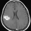
We report here on an uncommon case of metastatic choriocarcinoma to the lung, brain and lumbar spine. A 33-year-old woman was admitted to the pulmonary department with headache, dyspnea and hemoptysis. There was a history of cesarean section due to intrauterine fetal death at 37-weeks gestation and this occurred 2 weeks before admission to the pulmonary department. The radiological studies revealed a nodular lung mass with hypervascularity in the left upper lobe and also a brain parenchymal lesion in the parietal lobe with marginal bleeding and surrounding edema. She underwent embolization for the lung lesion, which was suspected to be an arteriovenous malformation according to the pulmonary arteriogram. Approximately 10 days after discharge from the pulmonary department, she was readmitted due to back pain and progressive paraparesis. The neuroradiological studies revealed a hypervascular tumor occupying the entire L3 vertebral body and pedicle, and the tumor extended to the epidural area. She underwent embolization of the hypervascular lesion of the lumbar spine, and after which injection of polymethylmethacrylate in the L3 vertebral body, total laminectomy of L3, subtotal removal of the epidural mass and screw fixation of L2 and L4 were performed. The result of biopsy was a choriocarcinoma.
Keywords: Metastatic choriocarconoma; Spinal metastasis.
Figures


Similar articles
-
Lumbar metastasis of choriocarcinoma.Spine (Phila Pa 1976). 2009 Jul 1;34(15):E538-43. doi: 10.1097/BRS.0b013e3181a98746. Spine (Phila Pa 1976). 2009. PMID: 19564760
-
Vertebral fracture after removing pedicle screws used for posterior lumbar interbody fusion: A case report.J Clin Neurosci. 2018 Nov;57:182-184. doi: 10.1016/j.jocn.2018.04.019. Epub 2018 Sep 19. J Clin Neurosci. 2018. PMID: 30243598
-
Integrated treatment of a lumbar vertebral hemangioma with spinal stenosis and radiculopathy: A case report and a review of the literature.J Craniovertebr Junction Spine. 2019 Oct-Dec;10(4):259-262. doi: 10.4103/jcvjs.JCVJS_106_19. Epub 2020 Jan 23. J Craniovertebr Junction Spine. 2019. PMID: 32089622 Free PMC article.
-
Low back pain in a child associated with acute onset cauda equina syndrome: a rare presentation of an aggressive vertebral hemangioma: a case report.J Pediatr Orthop. 2012 Apr-May;32(3):271-6. doi: 10.1097/BPO.0b013e318247195a. J Pediatr Orthop. 2012. PMID: 22411333 Review.
-
Paraneoplastic Encephalopathy in a Patient With Metastatic Lung Cancer: A Case Study.J Adv Pract Oncol. 2018 Mar;9(2):216-221. Epub 2018 Mar 1. J Adv Pract Oncol. 2018. PMID: 30588355 Free PMC article. Review.
Cited by
-
Metastatic choriocarcinoma to the lumbar spine: Case report and review of literature.Surg Neurol Int. 2014 Nov 21;5:161. doi: 10.4103/2152-7806.145205. eCollection 2014. Surg Neurol Int. 2014. PMID: 25525554 Free PMC article.
-
Intramedullary spinal cord metastasis of choriocarcinoma.J Korean Neurosurg Soc. 2012 Mar;51(3):141-3. doi: 10.3340/jkns.2012.51.3.141. Epub 2012 Mar 31. J Korean Neurosurg Soc. 2012. PMID: 22639709 Free PMC article.
-
Choriocarcinoma with multiple lung, skull and skin metastases in a postmenopausal female: A case report.Oncol Lett. 2015 Dec;10(6):3837-3839. doi: 10.3892/ol.2015.3750. Epub 2015 Sep 25. Oncol Lett. 2015. PMID: 26788218 Free PMC article.
-
Multidisciplinary treatment of life-threatening hemoptysis and paraplegia of choriocarcinoma with pulmonary, hepatic and spinal metastases: A case report.World J Clin Cases. 2020 Sep 6;8(17):3867-3874. doi: 10.12998/wjcc.v8.i17.3867. World J Clin Cases. 2020. PMID: 32953866 Free PMC article.
-
Choriocarcinoma presenting as an isolated bone marrow metastasis-a case report.Ecancermedicalscience. 2014 Jan 29;8:393. doi: 10.3332/ecancer.2014.393. eCollection 2014. Ecancermedicalscience. 2014. PMID: 24605133 Free PMC article.
References
-
- Allen SD, Lim AK, Seckl MJ, Blunt DM, Mitchell AW. Radiology of gestational trophoblastic neoplasia. Clin Radiol. 2006;61:301–313. - PubMed
-
- Baik EJ, Jung JE, Son WI, Song JC, Kim MR, Jung DY, et al. Clinical analysis of brain metastasis of choriocarcinoma. J Korean Cancer Assoc. 1993;25:673–679.
-
- Bas T, Aparisi F, Bas JL. Efficacy and safety of ethanol injection in 18 cases of vertebral hemangioma : a mean follow-up of 2 years. Spine (Phila Pa 1976) 2001;26:1577–1582. - PubMed
-
- Beskonakli E, Cayli S, Kulacoglu S. Metastatic choriocarcinoma in the thoracic extradural space : case report. Spinal Cord. 1998;36:366–367. - PubMed
-
- Chen HI, Heuer GG, Zaghloul K, Simon SL, Weigele JB, Grady MS. Lumbar vertebral hemangioma presenting with the acute onset of neurological symptoms. Case report. J Neurosurg Spine. 2007;7:80–85. - PubMed
Publication types
LinkOut - more resources
Full Text Sources

