

Does size matter? Comparison study between MRI, gross, and microscopic tumor sizes in breast cancer in lumpectomy specimens
- PMID: 20224728
- PMCID: PMC2836507
Does size matter? Comparison study between MRI, gross, and microscopic tumor sizes in breast cancer in lumpectomy specimens
Abstract
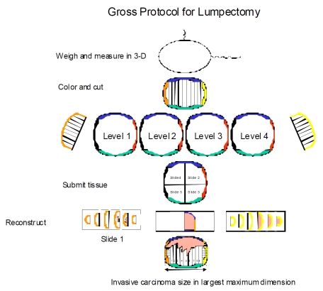
Size of breast cancer is essential in staging cancer to determine type and extent of patient management. This study was conducted to assess accuracy in estimating tumor size by MRI and gross using microscopy as gold standard. A retrospective study was done on 33 patients, 30-75 years, who underwent MRI of breasts with subsequent lumpectomy, 2002-2006, for invasive breast cancer. Size of lesion(s) on MRI and gross were compared with histological size. Of 37 lesions, 27 (73%) were invasive ductal (IDC) and 10 (27%) invasive lobular carcinoma (ILC). Tumor size by MRI matched histological size in 3%, underestimated 27%, and overestimated 70% of cases. Tumor size by gross matched histological size in 22%, underestimated 57%, and overestimated 22% of cases. MRI as an imaging modality and gross pathology both have significant limitations in measuring tumor size particularly in cases of invasive breast carcinoma. Random sectioning of lumpectomy specimen in invasive breast carcinoma may result in inaccurate staging of tumor by leading to false impression of tumor size and multi-focality and/or multi-centricity of tumor particularly in cases of ILC. Microscopic measurements of tumor size are necessary for accurate T-staging and recommended for appropriate patient management.
Keywords: Breast cancer; MRI; gross pathology; microscopic pathology; tumor size.
Figures
Comment in
-
Re: "Behjatnia B et al. "Does size matter? Comparison study between MRI, gross, and microscopic tumor sizes in breast cancer in lumpectomy specimens". IntJ Clin Exp Pathol 2010;3(3):303-309".Int J Clin Exp Pathol. 2010 Apr 30;3(4):458-60. Int J Clin Exp Pathol. 2010. PMID: 20490337 Free PMC article. No abstract available.
Similar articles
-
Breast magnetic resonance imaging for preoperative locoregional staging.Am J Surg. 2008 Sep;196(3):389-97. doi: 10.1016/j.amjsurg.2007.10.009. Epub 2008 Apr 23. Am J Surg. 2008. PMID: 18436185
-
Magnetic resonance imaging of breast cancer: factors affecting the accuracy of preoperative lesion sizing.Acta Radiol. 2015 Mar;56(3):260-8. doi: 10.1177/0284185114524089. Epub 2014 Feb 13. Acta Radiol. 2015. PMID: 24526754
-
Preliminary results: double lumpectomies for multicentric breast carcinoma.Am Surg. 2012 Dec;78(12):1345-8. Am Surg. 2012. PMID: 23265123
-
Mammography, sonography and MRI for detection and characterization of invasive lobular carcinoma of the breast.Breast Dis. 2008-2009;30:21-30. doi: 10.3233/BD-2009-0279. Breast Dis. 2008. PMID: 19850992 Review.
-
Complexities and challenges in the pathologic assessment of size (T) of invasive breast carcinoma.Adv Anat Pathol. 2014 Nov;21(6):420-32. doi: 10.1097/PAP.0000000000000040. Adv Anat Pathol. 2014. PMID: 25299311 Review.
Cited by
-
Preoperative imaging accuracy in size determination of prostate cancer in men undergoing radical prostatectomy for clinically localised disease.Insights Imaging. 2023 Jun 7;14(1):105. doi: 10.1186/s13244-023-01450-5. Insights Imaging. 2023. PMID: 37286770 Free PMC article.
-
Parathyroid weight estimation: beyond ellipsoid volume.Endocrine. 2025 Aug;89(2):587-594. doi: 10.1007/s12020-025-04273-0. Epub 2025 May 15. Endocrine. 2025. PMID: 40375055
-
Role of MRI in the staging of breast cancer patients: does histological type and molecular subtype matter?Br J Radiol. 2015;88(1055):20150458. doi: 10.1259/bjr.20150458. Epub 2015 Sep 16. Br J Radiol. 2015. PMID: 26374470 Free PMC article.
-
Deep learning in MRI-guided radiation therapy: A systematic review.J Appl Clin Med Phys. 2024 Feb;25(2):e14155. doi: 10.1002/acm2.14155. Epub 2023 Sep 15. J Appl Clin Med Phys. 2024. PMID: 37712893 Free PMC article.
-
Topographic enhancement mapping of the cancer-associated breast stroma using breast MRI.Integr Biol (Camb). 2011 Apr;3(4):490-6. doi: 10.1039/c0ib00089b. Epub 2011 Mar 18. Integr Biol (Camb). 2011. PMID: 21416100 Free PMC article.
References
-
- Greene FL, Page DL, Fleming ID, Firtz AG, Balch CM, Haller DG, Marrow M, et al., editors. 6th ed. New York: Springer; 2002. American Joint Committee on Cancer (AJCC) Cancer Staging Manual; pp. 221–40.
-
- Sobin LH, Wittekind CH. 6th edn. New York: Wiley; 2002. TNM Classification of Malignant Tumors.
-
- Fitzgibbons PL, Page DL, Weaver D, Thor AD, Allred DC, Clark GM, et al. Prognostic factors in breast cancer: College of American Pathologists consensus statement 1999. Arch Pathol Lab Med. 2000;124:966–78. - PubMed
-
- Pritt B, Ashikaga T, Oppenheimer RG, Weaver DL. Influence of breast cancer histology on the relationship between ultrasound and pathology tumor size measurements. Mod Pathol. 2004;17(8):905–10. - PubMed
-
- Apple SK, Suthar F. How do we measure a residual tumor size in istopathology (gold standard) after neoadjuvant chemotherapy? Breast. 2005;15(3):370–6. - PubMed