Development of acute lympboblastic leukemia in a patient with increased hematogones after toxic bone marrow damage
- PMID: 20224729
- PMCID: PMC2836508
Development of acute lympboblastic leukemia in a patient with increased hematogones after toxic bone marrow damage
Abstract
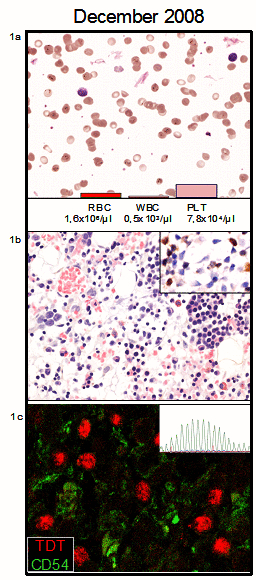
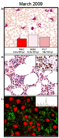
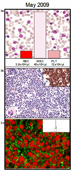
To the best of our knowledge we describe the first case showing the association of increased B cell precursors/hematogones in a regenerating post-toxic bone marrow with subsequent development of a B-ALL. Since all immunohistochemical/moleculargenetic anaylses have failed to identify the initial malignant leukemic clone, we suggest a close-meshed follow-up of such cases to identify potential mechanisms for the malignant transformation of B-cell precursors/hematogones and to prevent further fatal courses.
Keywords: B-ALL; Bone marrow; hematogones; toxic damage.
Figures


Similar articles
-
The knowledge on the 3rd type hematogones could contribute to more precise detection of small numbers of precursor B-acute lymphoblastic leukemia.Neoplasma. 2005;52(6):502-9. Neoplasma. 2005. PMID: 16284697
-
Bone marrow necrosis associated with the use of imatinib mesylate in a patient with Philadelphia chromosome-positive acute lymphoblastic leukemia.Ann Hematol. 2006 Aug;85(8):542-4. doi: 10.1007/s00277-005-0071-3. Epub 2006 Mar 29. Ann Hematol. 2006. PMID: 16570152 No abstract available.
-
[Histopathologic pattern of hyperplasia of bone marrow hematogones (medullar b lymphoid cell precursors) occurring after treatment of idiopathic myelofibrosis].Ann Pathol. 2008 Feb;28(1):27-31. doi: 10.1016/j.annpat.2007.10.001. Epub 2008 May 13. Ann Pathol. 2008. PMID: 18538711 French.
-
[Pediatric acute lymphoblastic leukemia initially presenting with bone marrow necrosis].Rinsho Ketsueki. 2007 Feb;48(2):140-3. Rinsho Ketsueki. 2007. PMID: 17370642 Review. Japanese.
-
Presentation and outcomes for children with bone marrow necrosis and acute lymphoblastic leukemia: a literature review.J Pediatr Hematol Oncol. 2011 Oct;33(7):e316-9. doi: 10.1097/MPH.0b013e318223fe9b. J Pediatr Hematol Oncol. 2011. PMID: 21941136 Review.
References
-
- Hassanein NM, Alcancia F, Perkinson KR, Buckley PJ, Lagoo AS. Distinct expression patterns of CD123 and CD34 on normal bone marrow B-cell precursors ("hematogones") and B Iymphobiastic leukemia blasts. Am J Clin Pathol. 2009;132:573–580. - PubMed
-
- Rimsza LM, Larson RS, Winter SS, et al. Benign hematogone-rich lymphoid proliferations can be distinguished from B-lineage acute Iymphobiastic leukemia by integration of morphology, im-munophenotype, adhesion molecule expression, and architectural features. Am J Clin Pathol. 2000;114:66–75. - PubMed
-
- Hendriks RW, Kersseboom R. Involvement of SLP-65 and Btk in tumor suppression and malignant transformation of pre-B cells. Semin Immunol. 2006;18:67–76. - PubMed
-
- Teitell MA, Pandolfi PP. Molecular genetics of acute Iymphobiastic leukemia. Annu Rev Pathol. 2009;4:175–198. - PubMed
Publication types
MeSH terms
Substances
LinkOut - more resources
Full Text Sources
Medical
