

Reactivation of latent tuberculosis in cynomolgus macaques infected with SIV is associated with early peripheral T cell depletion and not virus load
- PMID: 20224771
- PMCID: PMC2835744
- DOI: 10.1371/journal.pone.0009611
Reactivation of latent tuberculosis in cynomolgus macaques infected with SIV is associated with early peripheral T cell depletion and not virus load
Erratum in
-
Correction: reactivation of latent tuberculosis in cynomolgus macaques infected with SIV is associated with early peripheral T cell depletion and not virus load.PLoS One. 2015 Apr 13;10(4):e0124221. doi: 10.1371/journal.pone.0124221. eCollection 2015. PLoS One. 2015. PMID: 25874371 Free PMC article. No abstract available.
Abstract
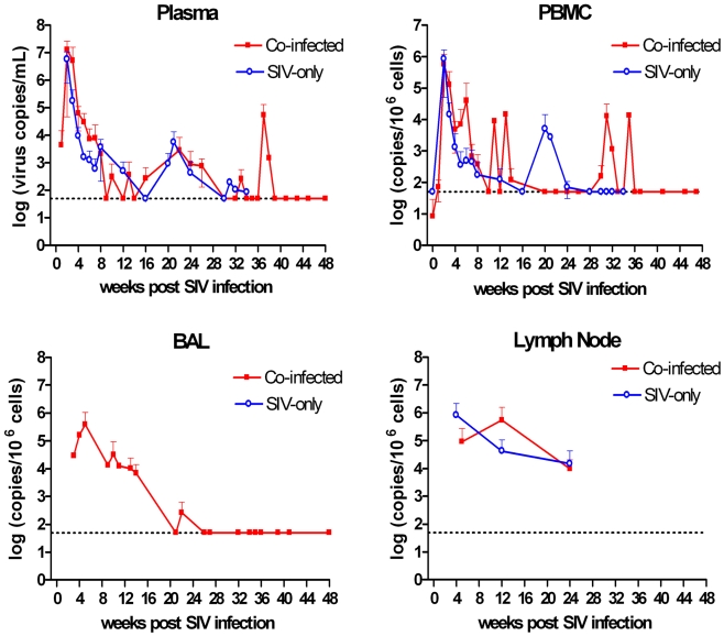
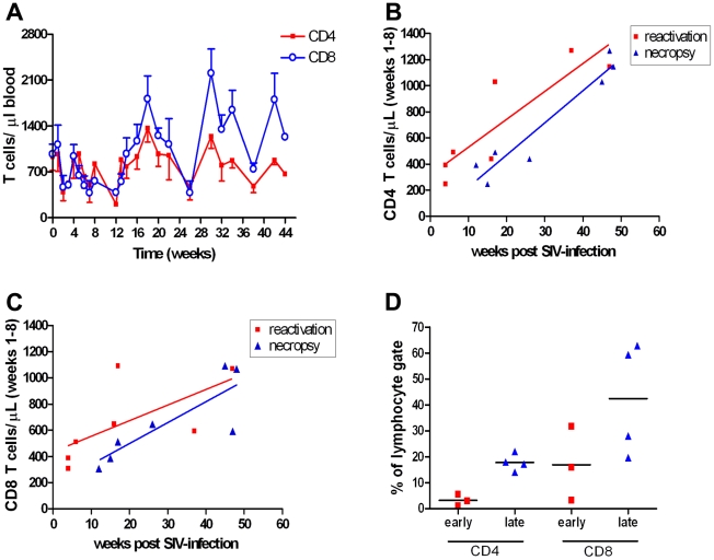
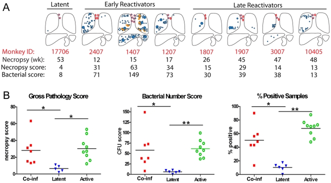
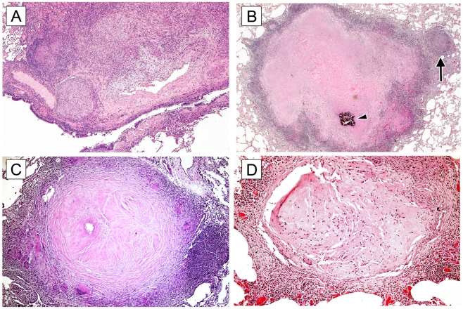
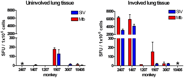
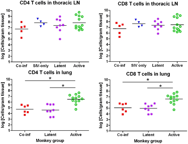
HIV-infected individuals with latent Mycobacterium tuberculosis (Mtb) infection are at significantly greater risk of reactivation tuberculosis (TB) than HIV-negative individuals with latent TB, even while CD4 T cell numbers are well preserved. Factors underlying high rates of reactivation are poorly understood and investigative tools are limited. We used cynomolgus macaques with latent TB co-infected with SIVmac251 to develop the first animal model of reactivated TB in HIV-infected humans to better explore these factors. All latent animals developed reactivated TB following SIV infection, with a variable time to reactivation (up to 11 months post-SIV). Reactivation was independent of virus load but correlated with depletion of peripheral T cells during acute SIV infection. Animals experiencing reactivation early after SIV infection (<17 weeks) had fewer CD4 T cells in the periphery and airways than animals reactivating in later phases of SIV infection. Co-infected animals had fewer T cells in involved lungs than SIV-negative animals with active TB despite similar T cell numbers in draining lymph nodes. Granulomas from these animals demonstrated histopathologic characteristics consistent with a chronically active disease process. These results suggest initial T cell depletion may strongly influence outcomes of HIV-Mtb co-infection.
Conflict of interest statement
Figures

Similar articles
-
Simian immunodeficiency virus-induced changes in T cell cytokine responses in cynomolgus macaques with latent Mycobacterium tuberculosis infection are associated with timing of reactivation.J Immunol. 2011 Mar 15;186(6):3527-37. doi: 10.4049/jimmunol.1003773. Epub 2011 Feb 11. J Immunol. 2011. PMID: 21317393 Free PMC article.
-
SIV and Mycobacterium tuberculosis synergy within the granuloma accelerates the reactivation pattern of latent tuberculosis.PLoS Pathog. 2020 Jul 30;16(7):e1008413. doi: 10.1371/journal.ppat.1008413. eCollection 2020 Jul. PLoS Pathog. 2020. PMID: 32730321 Free PMC article.
-
Spontaneous Control of SIV Replication Does Not Prevent T Cell Dysregulation and Bacterial Dissemination in Animals Co-Infected with M. tuberculosis.Microbiol Spectr. 2022 Jun 29;10(3):e0172421. doi: 10.1128/spectrum.01724-21. Epub 2022 Apr 25. Microbiol Spectr. 2022. PMID: 35467372 Free PMC article.
-
Chronic Immune Activation in TB/HIV Co-infection.Trends Microbiol. 2020 Aug;28(8):619-632. doi: 10.1016/j.tim.2020.03.015. Epub 2020 Apr 22. Trends Microbiol. 2020. PMID: 32417227 Free PMC article. Review.
-
Brain macrophages harbor latent, infectious simian immunodeficiency virus.AIDS. 2019 Dec 1;33 Suppl 2(Suppl 2):S181-S188. doi: 10.1097/QAD.0000000000002269. AIDS. 2019. PMID: 31789817 Free PMC article. Review.
Cited by
-
An Inflammatory Story: Antibodies in Tuberculosis Comorbidities.Front Immunol. 2019 Dec 9;10:2846. doi: 10.3389/fimmu.2019.02846. eCollection 2019. Front Immunol. 2019. PMID: 31921122 Free PMC article. Review.
-
Comparing adjuvanted H28 and modified vaccinia virus ankara expressingH28 in a mouse and a non-human primate tuberculosis model.PLoS One. 2013 Aug 19;8(8):e72185. doi: 10.1371/journal.pone.0072185. eCollection 2013. PLoS One. 2013. PMID: 23977248 Free PMC article.
-
Antiretroviral therapy timing impacts latent tuberculosis infection reactivation in a Mycobacterium tuberculosis/SIV coinfection model.J Clin Invest. 2022 Feb 1;132(3):e153090. doi: 10.1172/JCI153090. J Clin Invest. 2022. PMID: 34855621 Free PMC article.
-
SIV Infection Facilitates Mycobacterium tuberculosis Infection of Rhesus Macaques.Front Microbiol. 2017 Jan 13;7:2174. doi: 10.3389/fmicb.2016.02174. eCollection 2016. Front Microbiol. 2017. PMID: 28133458 Free PMC article.
-
Heterogeneity in tuberculosis.Nat Rev Immunol. 2017 Nov;17(11):691-702. doi: 10.1038/nri.2017.69. Epub 2017 Jul 24. Nat Rev Immunol. 2017. PMID: 28736436 Free PMC article. Review.
References
-
- Ulrichs T, Kosmiadi GA, Jorg S, Pradl L, Titukhina M, et al. Differential organization of the local immune response in patients with active cavitary tuberculosis or with nonprogressive tuberculoma. J Infect Dis. 2005;192:89–97. - PubMed
-
- Harries AD, Dye C. Tuberculosis. Ann Trop Med Parasitol. 2006;100:415–431. - PubMed
-
- Corbett EL, Watt CJ, Walker N, Maher D, Williams BG, et al. The growing burden of tuberculosis: global trends and interactions with the HIV epidemic. Arch Intern Med. 2003;163:1009–1021. - PubMed
-
- Post FA, Wood R, Pillay GP. Pulmonary tuberculosis in HIV infection: radiographic appearance is related to CD4+ T-lymphocyte count. Tuber Lung Dis. 1995;76:518–521. - PubMed
-
- Mukadi Y, Perriens JH, St Louis ME, Brown C, Prignot J, et al. Spectrum of immunodeficiency in HIV-1-infected patients with pulmonary tuberculosis in Zaire. Lancet. 1993;342:143–146. - PubMed
Publication types
MeSH terms
Substances
Grants and funding
LinkOut - more resources
Full Text Sources
Research Materials
