. 2010 Mar 10;5(3):e9620.
doi: 10.1371/journal.pone.0009620.

Morphological and genomic characterization of Filobasidiella depauperata: a homothallic sibling species of the pathogenic cryptococcus species complex

Affiliations

Morphological and genomic characterization of Filobasidiella depauperata: a homothallic sibling species of the pathogenic cryptococcus species complex

Marianela Rodriguez-Carres et al. PLoS One. .

Abstract

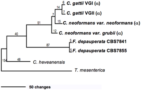
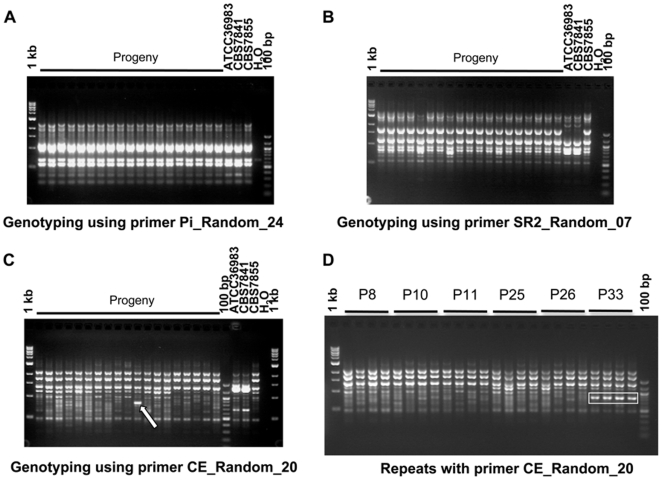
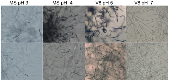
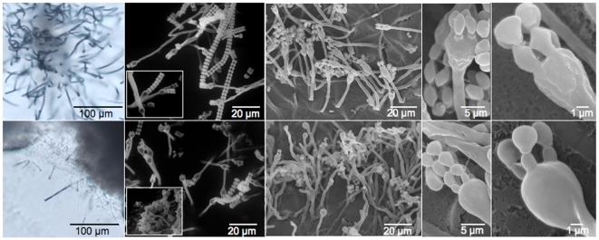
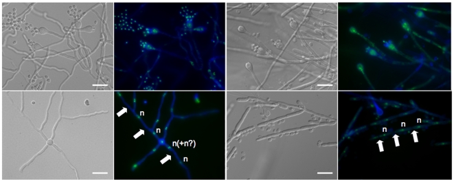
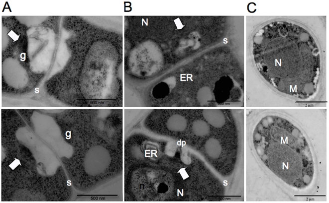
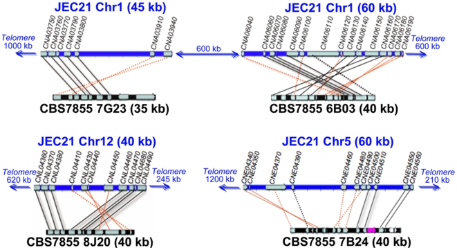
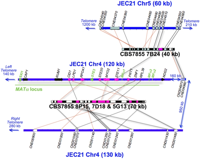
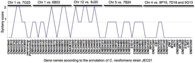
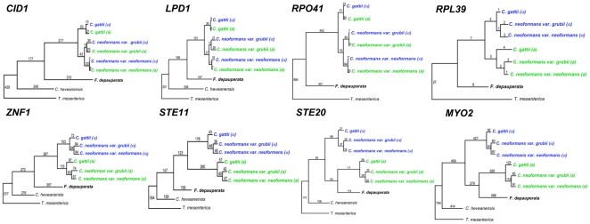
The fungal species Cryptococcus neoformans and Cryptococcus gattii cause respiratory and neurological disease in animals and humans following inhalation of basidiospores or desiccated yeast cells from the environment. Sexual reproduction in C. neoformans and C. gattii is controlled by a bipolar system in which a single mating type locus (MAT) specifies compatibility. These two species are dimorphic, growing as yeast in the asexual stage, and producing hyphae, basidia, and basidiospores during the sexual stage. In contrast, Filobasidiella depauperata, one of the closest related species, grows exclusively as hyphae and it is found in association with decaying insects. Examination of two available strains of F. depauperata showed that the life cycle of this fungal species shares features associated with the unisexual or same-sex mating cycle in C. neoformans. Therefore, F. depauperata may represent a homothallic and possibly an obligately sexual fungal species. RAPD genotyping of 39 randomly isolated progeny from isolate CBS7855 revealed a new genotype pattern in one of the isolated basidiospores progeny, therefore suggesting that the homothallic cycle in F. depauperata could lead to the emergence of new genotypes. Phylogenetic analyses of genes linked to MAT in C. neoformans indicated that two of these genes in F. depauperata, MYO2 and STE20, appear to form a monophyletic clade with the MATa alleles of C. neoformans and C. gattii, and thus these genes may have been recruited to the MAT locus before F. depauperata diverged. Furthermore, the ancestral MATa locus may have undergone accelerated evolution prior to the divergence of the pathogenic Cryptococcus species since several of the genes linked to the MATa locus appear to have a higher number of changes and substitutions than their MATalpha counterparts. Synteny analyses between C. neoformans and F. depauperata showed that genomic regions on other chromosomes displayed conserved gene order. In contrast, the genes linked to the MAT locus of C. neoformans showed a higher number of chromosomal translocations in the genome of F. depauperata. We therefore propose that chromosomal rearrangements appear to be a major force driving speciation and sexual divergence in these closely related pathogenic and saprobic species.


Conflict of interest statement

Competing Interests: The authors have declared that no competing interests exist.

Figures

Figure 1
Figure 1. Phylogeny of F. depauperata and closely related species.
A Maximum Parsimony tree depicting the relationship between both strains of F. depauperata and the pathogenic Cryptococcus species using a concatenated data set derived from two coding genes. Numbers above the branches indicate changes. Statistical support was calculated from 1,000 bootstrap replicates. Bootstrap values were >70% in all branches (values not shown). The “α” indicates strains with the MATα locus. Unrooted trees for Maximum Parsimony and Maximum likelihood are shown in supporting File S1.
Figure 2
Figure 2. Genetic diversity between isolates of F. depauperata and among the progeny of isolate CBS7855.
Panels A, B and C shows representative results from three of the 12 different random primers that were used to evaluate the genetic similarity of both strains of F. depauperata, and progeny from CBS7855. Strains CBS7841 and ATCC36983 represent the same isolate, but obtained from two different culture collections (ATCC and CBS). A total of 39 progeny were tested, but only 19 are shown in panels A, B & C. Panel A shows the results from one of the random primers (Pi_Random_24) that generated an identical PCR pattern for all isolates (CBS7841, CBS7855, and also among the progeny from CBS7855). Panel B and C shows the results from the two random primers (SR2_Random_07, and CE_Random_20) that displayed different PCR patterns for CBS7841 and CBS7855. Primer CE_Random_20 also revealed a unique band (white arrow) present in progeny #33 (P33), but not present in the parental strain CBS7855. Panel D shows the consistency and reproducibility of the unique band observed in P33 by independent repetitions of the PCR experiments. Rectangles indicated the unique PCR band that is present P33 but absent from the parental strain and other progeny. P8, P10, P11, P25, P26, and P33 correspond to progeny of CBS7855. 1 kb∶1 kb DNA ladder; 100 bp∶100 bp DNA ladder; H2O: negative control for the PCR reaction.
Figure 3
Figure 3. Fruiting and filamentation of F. depauperata at different pHs.
Colony-edges (4× magnification) of F. depauperata growing on different types of mating media and at different pH (MS pH = 3, MS pH = 4, V8 pH = 5, and V8 pH = 7) are shown. Top panels are of strain CBS7841, and bottom panels strain CBS7855. Photographs were taken after 7 days.
Figure 4
Figure 4. Basidia and basidiospores morphology differ between the two strains of F. depauperata.
SEM images show that CBS7841 basidia have a rough texture, while the basidia of CBS7855 are smooth. Longer spore chains were also observed on the basidia of CBS7841. Surface field views of the colony-edges were examined by SEM. Top panel shows strain CBS7841 and bottom panel CBS7855.
Figure 5
Figure 5. Monokaryotic hyphae, basidia, and basidiospores of F. depauperata.
Hyphae and basidiospores from both strains of F. depauperata appear to be mostly monokaryotic with hyphae that lack clamp connections. Nuclear content was determined by examination under fluorescence light microscopy (color images). Samples were stained with sytox-green to detect nucleic acids (shown in green), and with calcofluor white to visualize the cell walls (shown in blue). DIC images are shown in grey. White arrows indicate cell wall septa, and the letter “n” represents the observed nuclear content. The two left panels are of strain CBS7841, and the two right panels are CBS7855.
Figure 6
Figure 6. Transmission electron microscopy (TEM) of hyphal septa and basidiospores of F. depauperata.
Panels A and B show different TEM images of the surface view of dolipore septa which is characteristic of basidiomycetes. Panel C shows TEM images from spores. The top row shows TEM images from strain CBS7855 and the bottom row from CBS7841. Arrows indicate electron dense occlusions and vesicles associated with the single septal opening. N denotes the nucleous, M the mitochondria, n the nucleolus, ER the endoplasmic reticulum, dp the dolipore, g the granular dolipore plug, and S the septa. Top panel shows strain CBS7855, and bottom panel shows CBS7841.
Figure 7
Figure 7. Genomic comparison between C. neoformans and F. depauperata.
The gene order between F. depauperata and C. neoformans is conserved. Chromosomes of C. neoformans var. neoformans strain JEC21 are drawn in blue. Sequenced fosmids from the F. depauperata library of strain CBS7855 are drawn in black. Black lines connect genes in the same orientation, while red lines indicate inversions. Solid lines point to syntenic blocks where the adjacent gene order is conserved. Dotted lines connect orthologous genes where the adjacent genes are not conserved. Genes located on chromosomes 1, 5, and 12 of C. neoformans are shown in grey. The aim of this figure is to display the gene arrangements and directions. For simplicity purposes, only those genes present in both species, C. neoformans and F. depauperata, are shown.
Figure 8
Figure 8. Genomic comparison of MAT-associated genes in C. neoformans.
Several chromosomal translocations, fusions, and inversions appear be present in F. depauperata by comparison to the genome of C. neoformans. Chromosome 4 of C. neoformans var. neoformans strain JEC21 is drawn in blue. Sequenced fosmids from the F. depauperata library of strain CBS7855 are drawn in black. Black lines connect genes in the same orientation, while red lines indicate inversions. Solid lines point to syntenic blocks where the adjacent gene order is conserved. Dotted lines connect orthologous genes where the adjacent genes are not conserved. The aim of this figure is to display the gene arrangements and directions. For simplicity purposes, only those genes present in both species, C. neoformans and F. depauperata, are shown in grey, white, and pink. Genes located on chromosomes 5 and 12 of C. neoformans are shown in grey. Genes located on chromosome 4 of C. neoformans are shown in white while genes associated with the MAT locus of Cryptococcus are shown in pink. The α locus and the mating genes (the pheromone genes MFα, the pheromone receptor gene STE3, and the homeodomain transcription factor SXI1) of C. neoformans are shown in green. Genes shown in black, CAP1 and RUM1, are associated with the MAT locus of C. neoformans, and NOG2 flanks the MAT locus. These three genes were sequenced and identified in F. depauperata, however fosmids containing these genes were not found in the genomic library of F. depauperata.
Figure 9
Figure 9. Graphical display of synteny between F. depauperata and C. neoformans.
Summary of the synteny over the different genomic regions compared between F. depauperata and C. neoformans. The x-axis shows the gene names according to the annotations in the genome of C. neoformans strains JEC21. The y-axis shows the level of synteny. The maximum synteny score (score  = 2) was assigned to those ORFs fully syntenic because they are located between two adjacently conserved genes, while ORFs of adjacent pairs were assigned a lower score (score  = 1). Those ORFs with no adjacent genes conserved between the two species were given the lowest synteny score (score  = 0). The scoring system disregards the direction in which the genes are transcribed, and focuses on gene order by displaying the score for each gene calculated from the number of syntenic adjacent genes.
Figure 10
Figure 10. Phylogeny of genes linked to the MAT locus of C. neoformans.
Two of the genes from F. depauperata, STE20 and MYO2, hypothesized to be ancestrally acquired in the pheromone/pheromone receptor locus of the pathogenic Cryptococcus species, clustered with the STE20 a and MYO2 a alleles of C. gattii and C. neoformans. The remaining genes from F. depauperata exhibit a species-specific phylogeny. Genes from the MAT a alleles are shown in green, and genes from the MATα allele are shown in blue. (α) indicates strains with the MATα locus, and (a) indicates strains with the MAT a locus. The tree was constructed using Maximum Parsimony, and numbers indicate total changes in the branches. Statistical support was calculated from 1,000 bootstrap replicates. Bootstrap values were >70% in all branches (values not shown). Unrooted trees for Maximum Parsimony and Maximum likelihood are shown in supporting File S1.

Similar articles

Cited by

References

    1. Hurst LD, Pál CP, Lercher MJ. The evolutionary dynamics of eukaryotic gene order. Nature Reviews. 2004;5:299–310. - PubMed
    1. Fraser JA, Diezmann S, Subaran RL, Allen A, Lengeler KB, et al. Convergent evolution of chromosomal sex-determining regions in the animal and fungal kingdoms. PLoS Biology. 2004;2:2243–2255. - PMC - PubMed
    1. Casselton LA, Olesnicky NS. Molecular genetics of mating recognition in basidiomycete fungi. Microbiol Mol Biol Rev. 1999;62:55–70. - PMC - PubMed
    1. Kahmann RR, Romeis T, Bolker M, Kamper J. Control of mating and development in Ustilago maydis. Curr Opin Genet Dev. 1995;5:559–564. - PubMed
    1. Kronstad JW, Staben C. Mating type in filamentous fungi. Annu Rev Genet. 1997;31:245–276. - PubMed

Publication types

LinkOut - more resources