

Unique biological properties of catalytic domain directed human anti-CAIX antibodies discovered through phage-display technology
- PMID: 20224781
- PMCID: PMC2835754
- DOI: 10.1371/journal.pone.0009625
Unique biological properties of catalytic domain directed human anti-CAIX antibodies discovered through phage-display technology
Abstract
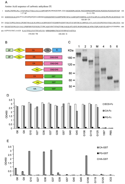
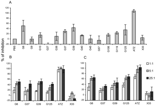
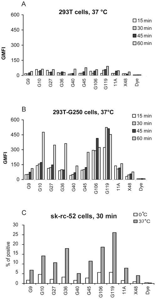
Carbonic anhydrase IX (CAIX, gene G250/MN-encoded transmembrane protein) is highly expressed in various human epithelial tumors such as renal clear cell carcinoma (RCC), but absent from the corresponding normal tissues. Besides the CA signal transduction activity, CAIX may serve as a biomarker in early stages of oncogenesis and also as a reliable marker of hypoxia, which is associated with tumor resistance to chemotherapy and radiotherapy. Although results from preclinical and clinical studies have shown CAIX as a promising target for detection and therapy for RCC, only a limited number of murine monoclonal antibodies (mAbs) and one humanized mAb are available for clinical testing and development. In this study, paramagnetic proteoliposomes of CAIX (CAIX-PMPLs) were constructed and used for anti-CAIX antibody selection from our 27 billion human single-chain antibody (scFv) phage display libraries. A panel of thirteen human scFvs that specifically recognize CAIX expressed on cell surface was identified, epitope mapped primarily to the CA domain, and affinity-binding constants (KD) determined. These human anti-CAIX mAbs are diverse in their functions including induction of surface CAIX internalization into endosomes and inhibition of the carbonic anhydrase activity, the latter being a unique feature that has not been previously reported for anti-CAIX antibodies. These human anti-CAIX antibodies are important reagents for development of new immunotherapies and diagnostic tools for RCC treatment as well as extending our knowledge on the basic structure-function relationships of the CAIX molecule.
Conflict of interest statement
Figures


Similar articles
-
Human anti-CAIX antibodies mediate immune cell inhibition of renal cell carcinoma in vitro and in a humanized mouse model in vivo.Mol Cancer. 2015 Jun 11;14:119. doi: 10.1186/s12943-015-0384-3. Mol Cancer. 2015. PMID: 26062742 Free PMC article.
-
Carbonic anhydrase IX in renal cell carcinoma: implications for prognosis, diagnosis, and therapy.Eur Urol. 2010 Jul;58(1):75-83. doi: 10.1016/j.eururo.2010.03.015. Epub 2010 Mar 23. Eur Urol. 2010. PMID: 20359812 Review.
-
Binding of the phage display derived peptide CaIX-P1 on human colorectal carcinoma cells correlates with the expression of carbonic anhydrase IX.Int J Mol Sci. 2012 Oct 11;13(10):13030-48. doi: 10.3390/ijms131013030. Int J Mol Sci. 2012. PMID: 23202936 Free PMC article.
-
A novel VHH nanobody against the active site (the CA domain) of tumor-associated, carbonic anhydrase isoform IX and its usefulness for cancer diagnosis.Biotechnol Lett. 2014 Jan;36(1):21-8. doi: 10.1007/s10529-013-1340-1. Epub 2013 Sep 26. Biotechnol Lett. 2014. PMID: 24068505
-
Carbonic anhydrase IX as an anticancer therapy target: preclinical evaluation of internalizing monoclonal antibody directed to catalytic domain.Curr Pharm Des. 2010;16(29):3255-63. doi: 10.2174/138161210793429832. Curr Pharm Des. 2010. PMID: 20819068 Review.
Cited by
-
Visualization and quantitation of the expression of microRNAs and their target genes in neuroblastoma single cells using imaging cytometry.BMC Res Notes. 2011 Nov 28;4:517. doi: 10.1186/1756-0500-4-517. BMC Res Notes. 2011. PMID: 22123030 Free PMC article.
-
Overcoming Hypoxia-Mediated Tumor Progression: Combinatorial Approaches Targeting pH Regulation, Angiogenesis and Immune Dysfunction.Front Cell Dev Biol. 2016 Mar 31;4:27. doi: 10.3389/fcell.2016.00027. eCollection 2016. Front Cell Dev Biol. 2016. PMID: 27066484 Free PMC article. Review.
-
Carbonic Anhydrase IX: A Renewed Target for Cancer Immunotherapy.Cancers (Basel). 2022 Mar 9;14(6):1392. doi: 10.3390/cancers14061392. Cancers (Basel). 2022. PMID: 35326544 Free PMC article. Review.
-
Format-tuning of in vivo-launched bispecific T cell engager enhances efficacy against renal cell carcinoma.J Immunother Cancer. 2024 Jun 4;12(6):e008733. doi: 10.1136/jitc-2023-008733. J Immunother Cancer. 2024. PMID: 38834201 Free PMC article.
-
Anti-CAIX BBζ CAR4/8 T cells exhibit superior efficacy in a ccRCC mouse model.Mol Ther Oncolytics. 2021 Dec 31;24:385-399. doi: 10.1016/j.omto.2021.12.019. eCollection 2022 Mar 17. Mol Ther Oncolytics. 2021. PMID: 35118195 Free PMC article.
References
-
- Amato RJ. Renal cell carcinoma: review of novel single-agent therapeutics and combination regimens. Ann Oncol. 2005;16:7–15. - PubMed
-
- Cohen HT, McGovern FJ. Renal-cell carcinoma. N Engl J Med. 2005;353:2477–2490. - PubMed
-
- Motzer RJ, Mazumdar M, Bacik J, Russo P, Berg WJ, et al. Effect of cytokine therapy on survival for patients with advanced renal cell carcinoma. J Clin Oncol. 2000;18:1928–1935. - PubMed
-
- Motzer RJ, Russo P. Systemic therapy for renal cell carcinoma. J Urol. 2000;163:408–417. - PubMed
Publication types
MeSH terms
Substances
Grants and funding
LinkOut - more resources
Full Text Sources
Other Literature Sources
Molecular Biology Databases
